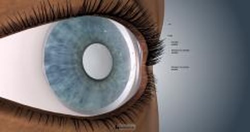
Foreign bodies in the cornea cause abrasions, resulting in pain and redness, and lead to infections, even after they are removed. Most of these injuries are minor.
(See also Overview of Eye Injuries.)
The most common injuries involving the surface of the transparent dome on the front surface of the eye (cornea) are
Scratches (abrasions)
Foreign bodies (objects)
About a quarter of people who present to an emergency department complaining of eye pain have a corneal abrasion.
Causes of Corneal Abrasions and Corneal Foreign Bodies
Particles are common causes of corneal abrasions. Particles can be dispersed via explosions, wind, or working with tools (for example, grinders, saws, hammers, drills, or rotary tools with a metal-on-metal mechanism). Contact lenses are a common cause of corneal abrasions. Poorly fitting lenses, lenses worn when the eyes are dry, lenses that have been incompletely cleaned and that have particles attached to them, lenses left in the eyes too long, lenses left in inappropriately during sleep, and forceful or inept removal of lenses can result in scratches on the surface of the eyes. Other common sources of abrasions are
Tree branches or falling debris
Fingernails
Hairbrushes
Make-up applicators
Most corneal abrasions heal without developing infections (such as conjunctivitis and corneal ulcers), but those related to contact lenses or contaminated with soil or vegetable matter (for example, an injury caused by a tree branch) are more likely to become infected.
Symptoms of Corneal Abrasions and Corneal Foreign Bodies
Corneal abrasions and foreign bodies usually cause pain, tearing, and a feeling that there is something in the eye. They may also cause redness (due to dilated blood vessels on the surface of the eye) or, occasionally, swelling of the eye and eyelid. Vision may become blurred. Light may cause the muscle that constricts the pupil to undergo a painful spasm.
Injuries that penetrate the eye (intraocular foreign bodies) may cause similar symptoms. If a foreign object penetrates the inside of the eye, fluid may leak out.
Diagnosis of Corneal Abrasions and Corneal Foreign Bodies
A doctor's evaluation
Prompt diagnosis and appropriate treatment of corneal abrasions and foreign bodies can help prevent infection of the cornea (corneal ulcer), infection of the inside of the eye (endophthalmitis) or inflammation of the iris (iridocyclitis), all of which put vision at risk. The diagnosis is based on the person’s symptoms, the circumstances of the injury, and the examination.
Treatment of Corneal Abrasions and Corneal Foreign Bodies
Removal of foreign bodies
Antibiotics
Pain relief
Corneal foreign bodies
Prior to removing a corneal foreign body, the doctor usually numbs the surface of the eye with an anesthetic drop (such as proparacaine). The doctor also administers an eye drop containing a dye (fluorescein) that glows under special lighting, making surface objects more visible and revealing abrasions. Using a Prior to removing a corneal foreign body, the doctor usually numbs the surface of the eye with an anesthetic drop (such as proparacaine). The doctor also administers an eye drop containing a dye (fluorescein) that glows under special lighting, making surface objects more visible and revealing abrasions. Using aslit lamp or other magnifying instrument, the doctor then removes any remaining foreign objects. Often the foreign object can be lifted out with a moist sterile cotton swab or flushed out with sterile water (irrigation). If the person is able to stare without moving the eye, foreign objects that cannot be dislodged easily with a swab can often be removed painlessly with a sterile hypodermic needle or a special instrument.
When iron or steel foreign bodies are removed, they can leave a ring of rust, which may need to be removed with a sterile hypodermic needle or a low-speed rotary sterile burr (a small surgical tool with a tiny, rotating, grinding, and drilling surface).
Sometimes a foreign body is trapped under the upper eyelid. The eyelid must be flipped over (a painless procedure called eversion) to remove the foreign body. Doctors may also gently rub a sterile cotton swab over the inside of the eyelid to remove any tiny particles that may not be visible.
Corneal abrasions
Corneal abrasions are treated similarly whether or not a foreign body was removed. Usually, an antibiotic ointment (for example, bacitracin with polymyxin B) is given for a few days to prevent infection. Large abrasions may require additional treatment. The pupil is kept dilated with cycloplegic eye drops (such as cyclopentolate or homatropine) if people are sensitive to light. These drops prevent painful spasm of the muscles that constrict the pupil. Corneal abrasions are treated similarly whether or not a foreign body was removed. Usually, an antibiotic ointment (for example, bacitracin with polymyxin B) is given for a few days to prevent infection. Large abrasions may require additional treatment. The pupil is kept dilated with cycloplegic eye drops (such as cyclopentolate or homatropine) if people are sensitive to light. These drops prevent painful spasm of the muscles that constrict the pupil.
Pain can be treated with oral medications such as acetaminophen or occasionally prescription pain medication. Some doctors give diclofenac or ketorolac eye drops to help relieve pain, but care must be taken because these medications could rarely cause complications such as a type of corneal scarring (called corneal melting). Anesthetics that are applied directly to the eye, although they relieve pain effectively, should not be used after evaluation and treatment because they can impair healing. Pain can be treated with oral medications such as acetaminophen or occasionally prescription pain medication. Some doctors give diclofenac or ketorolac eye drops to help relieve pain, but care must be taken because these medications could rarely cause complications such as a type of corneal scarring (called corneal melting). Anesthetics that are applied directly to the eye, although they relieve pain effectively, should not be used after evaluation and treatment because they can impair healing.
Eye patches may increase the risk of infection and usually are not used, particularly for abrasions that result from a contact lens or an object that may be contaminated with soil or vegetable matter.
Prognosis for Corneal Abrasions and Corneal Foreign Bodies
Fortunately, the surface cells of the eye regenerate rapidly. Even large corneal abrasions tend to heal in 1 to 3 days. A contact lens should not be worn for 5 days after the abrasion heals. A follow-up examination by an ophthalmologist (a medical doctor who specializes in the evaluation and treatment—surgical and nonsurgical—of eye disorders) 1 or 2 days after the injury is wise, but the time frame may vary based on the size and severity of the injury.

