


(See also Overview of Fractures.)
The scaphoid is the most commonly injured carpal bone. Scaphoid fractures usually result from wrist hyperextension, typically during a fall on an outstretched hand. They can disrupt the blood supply to the proximal scaphoid. Osteonecrosis is thus a common complication, even when initial care is optimal, and can cause disabling, degenerative arthritis of the wrist.
Symptoms and Signs of Scaphoid Fractures
The radial side of the wrist is swollen and tender. If patients have these symptoms, scaphoid fracture should be considered. More specific signs include
Pain during axial compression of the thumb
Pain during wrist supination against resistance
Particularly tenderness in the anatomic snuffbox during ulnar wrist deviation
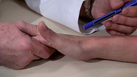
The anatomic snuffbox is palpated just distal to the radius between the extensor pollicis longus, extensor pollicis brevis, and abductor pollicis longus tendons.
Diagnosis of Scaphoid Fractures
Radiographs
MRI
If a scaphoid fracture is suspected and imaging is nondiagnostic, presumptive treatment with a thumb spica splint and follow-up radiograph
Initially, radiographs (anteroposterior, lateral, and oblique views) are taken but are often normal. Only around 70% of scaphoid fractures are seen on initial radiographs (1).
If radiographs are normal but a fracture is still suspected, MRI can be done. MRI may be used to diagnose scaphoid fractures because it is more accurate than CT or bone scanning in the acute setting (1).

ZEPHYR/SCIENCE PHOTO LIBRARY
If a fracture is suspected clinically and imaging is nondiagnostic, it is treated presumptively as a fracture and a thumb spica splint is applied. The patient should be re-examined 1 week after injury. If the patient is still in pain or if the wrist is tender when examined 1 week after injury, repeat radiographs, CT scan, or MRI should be obtained.

Pearls & Pitfalls
|
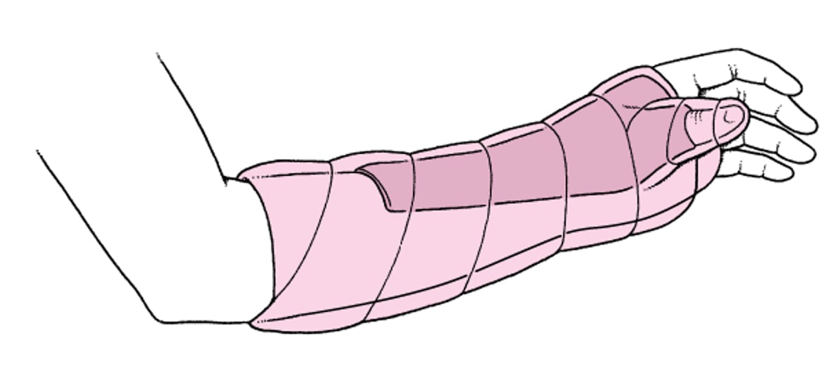
Thumb Spica Splint

Diagnosis references
1. Gäbler C, Kukla C, Breitenseher MJ, Trattnig S, Vécsei V. Diagnosis of occult scaphoid fractures and other wrist injuries. Are repeated clinical examinations and plain radiographs still state of the art?. Langenbecks Arch Surg. 2001;386(2):150-154. doi:10.1007/s004230000195
2. Carpenter CR, Pines JM, Schuur JD, et al. Adult scaphoid fracture. Acad Emerg Med. 21 (2):101–121, 2014. doi: 10.1111/acem.12317
Treatment of Scaphoid Fractures
Thumb spica immobilization
Many nondisplaced fractures can be treated definitively with a thumb spica cast, which is worn for up to 6 to 8 weeks. A thumb spica splint can be applied if cast application needs to be deferred.
Sometimes open reduction with internal fixation (ORIF) is required when there is moderate displacement of the fracture segments.
Key Points
Scaphoid fractures usually result from wrist hyperextension, typically during a fall on an outstretched hand.
These fractures can disrupt the blood supply to the proximal scaphoid; thus, osteonecrosis is a common, sometimes disabling, complication.
Take anteroposterior, lateral, and oblique radiographs; if imaging is normal or nondiagnostic but clinical findings suggest a scaphoid fracture, do MRI or immobilize with thumb spica splint and arrange for repeat radiographs in 1 week.


