


Craniosynostosis is premature fusion of 1 or more calvarial sutures (fibrous joints between bones of the skull).
(See also Overview of Congenital Craniofacial Anomalies.)
Premature fusion of sutures causes a characteristic skull deformity due to decreased growth in a direction perpendicular to the closed suture. It occurs in 1 of 2500 live births (1). There are several types, depending on which suture is fused.
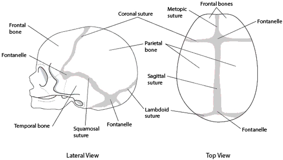
Skull (Calvarial) Sutures

A clinical geneticist should evaluate affected patients even in cases of apparent isolated congenital anomaly.
Chromosomal microarray analysis, specific gene tests, or broader gene panel tests should be considered in the evaluation of patients with congenital craniofacial anomalies. If the results of these tests are nondiagnostic, whole exome sequencing analysis may be recommended.
Reference
1. Boulet SL, Rasmussen SA, Honein MA. A population-based study of craniosynostosis in metropolitan Atlanta, 1989-2003. Am J Med Genet A. 2008;146A(8):984-991. doi:10.1002/ajmg.a.32208
Sagittal Craniosynostosis
Sagittal craniosynostosis is the most common type and causes a narrow and long skull (dolichocephaly). Most cases are isolated and sporadic, with recurrence risk of transmission to future offspring < 3% (1, 2).
Learning disability is common among affected patients, with reports ranging from 20 to 50% (1).
Several genes have been implicated in sagittal craniosynostosis, but chromosomal microarray analysis is not typically necessary unless developmental delays or other congenital anomalies are present.


Sagittal craniosynostosis references
1. Greenwood J, Flodman P, Osann K, Boyadjiev SA, Kimonis V. Familial incidence and associated symptoms in a population of individuals with nonsyndromic craniosynostosis. Genet Med. 2014;16(4):302-310. doi:10.1038/gim.2013.134
2. Lajeunie E, Le Merrer M, Bonaïti-Pellie C, Marchac D, Renier D. Genetic study of scaphocephaly. Am J Med Genet. 1996;62(3):282-285. doi:10.1002/(SICI)1096-8628(19960329)62:3<282::AID-AJMG15>3.0.CO;2-G
Coronal Craniosynostosis
Coronal craniosynostosis is the second most common type and can be bilateral, causing a short and broad skull (brachycephaly), or unilateral, causing a diagonal skull deformity (plagiocephaly). True plagiocephaly (ie, caused by craniosynostosis) often results in asymmetric orbits and is to be differentiated from positional plagiocephaly, which is due to torticollis or positioning the infant predominantly on one side and does not result in asymmetric orbits. In positional plagiocephaly, the back of the skull is flattened on one side, there is frontal bossing on the same side, and the ear on the flattened side may be pushed forward, but the orbits remain symmetrical.
About 25% of coronal craniosynostosis cases are syndromic and due to single-gene pathogenic variants or chromosomal defects (1). Pathogenic variants in several genes have been identified in patients with isolated nonsyndromic coronal craniosynostosis. Genetic diagnoses account for approximately 21% of all cases of craniosynostosis and are associated with increased rates of medical complications (2). Gene panel tests are currently recommended even in sporadic cases.
Coronal craniosynostosis is commonly associated with facial and extracranial anomalies that occur in numerous genetic syndromes, including Crouzon, Muenke, Pfeiffer, Saethre-Chotzen, Carpenter, and Apert syndromes. These syndromes can be confirmed by genetic testing.


Coronal craniosynostosis references
1. Lattanzi W, Barba M, Di Pietro L, Boyadjiev SA. Genetic advances in craniosynostosis. Am J Med Genet A. 2017;173(5):1406-1429. doi:10.1002/ajmg.a.38159
2. Wilkie AO, Byren JC, Hurst JA, et al. Prevalence and complications of single-gene and chromosomal disorders in craniosynostosis. Pediatrics. 2010;126(2):e391-e400. doi:10.1542/peds.2009-3491


