- Pregnancy Test and Due Date
- Stages of Fetal Development
- Physical Changes During Pregnancy
- Medical Care During Pregnancy
- Self-Care During Pregnancy
- Anemia During Pregnancy
- Infections During Pregnancy
- Blood Clot Disorders During Pregnancy
- Urinary Tract Infections During Pregnancy
- Multiple Pregnancy
- Late-Term and Postterm Pregnancy


A pregnancy goes through several stages of development. A fertilized egg develops into a blastocyst, then an embryo, then a fetus.
Fertilization
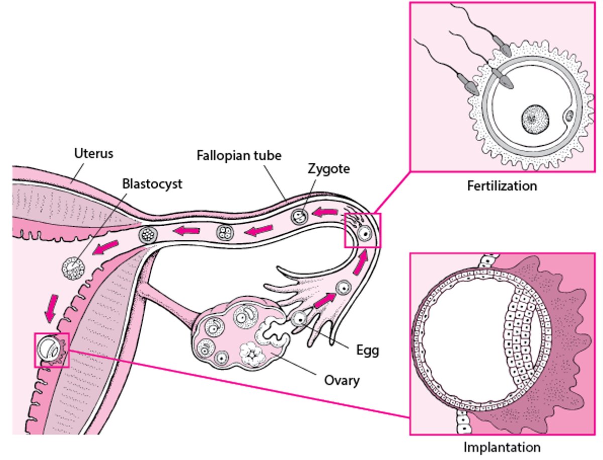
During each normal menstrual cycle, 1 egg (oocyte) is usually released from of the ovaries, about 14 days after the last menstrual period. Release of the egg is called ovulation. The egg then enters into the funnel-shaped end of 1 of the fallopian tubes.
At ovulation, the mucus in the cervix (bottom part of the uterus) becomes more fluid and more elastic, allowing sperm to enter the uterus rapidly. Within 5 minutes, sperm may move from the vagina, through the cervix into the uterus, and to the fallopian tube—the usual site of fertilization.
If fertilization does not occur, the egg moves through the fallopian tube into the uterus, and it is passed out of the uterus with the next menstrual period.
If a sperm penetrates the egg, fertilization occurs. Cells lining the fallopian tube have hair-like structures, called cilia, that help sweep the fertilized egg (zygote) through the tube and into the uterine cavity. The cells of the zygote divide (split into 2 cells) repeatedly as the zygote moves down the fallopian tube to the uterus. The zygote enters the uterus within 3 to 5 days.
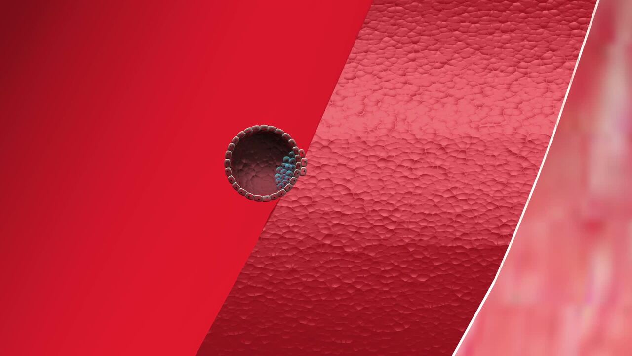
In the uterus, the cells continue to divide, becoming a hollow ball of cells called a blastocyst. The blastocyst implants in the wall of the uterus about 6 days after fertilization.
A twin pregnancy may occur in 2 different ways: identical or fraternal. Identical twins result when 1 fertilized egg separates into 2 embryos after it has begun to divide. Because 1 egg was fertilized by 1 sperm, the genetic material in the 2 embryos is the same. If more than 1 egg is released and fertilized, the resulting twins are fraternal rather than identical because the genetic material in each egg and in each sperm is slightly different.
In a triplet pregnancy, 3 eggs may be fertilized or, sometimes, 2 of the embryos are identical twins (resulting form 1 fertilized egg that divided into 2) and the third embryo is nonidentical. Different combinations of identical and non-identical embryos may also occur in pregnancies with even more than 3 embryos.
From Egg to Embryo
Once a month, an egg is released from an ovary into a fallopian tube. After sexual intercourse, sperm move from the vagina through the cervix into the uterus and then into the fallopian tubes, where 1 sperm fertilizes the egg. The fertilized egg (zygote) divides repeatedly as it moves down the fallopian tube to the uterus. First, the zygote becomes a solid ball of cells. Then it becomes a hollow ball of cells called a blastocyst. Inside the uterus, the blastocyst implants in the wall of the uterus, where it develops into an embryo attached to a placenta and surrounded by fluid-filled membranes. |

Development of the Blastocyst
About 6 days after fertilization, the blastocyst attaches to the wall of the uterine cavity, usually near the top. This process, called implantation, is completed by day 9 or 10.

The wall of the blastocyst is 1 cell thick except in 1 area, where it is 3 to 4 cells thick. The inner cells in the thickened area develop into the embryo, and the outer cells burrow into the wall of the uterus and develop into the placenta. The placenta produces several hormones that help maintain the pregnancy. For example, the placenta produces human chorionic gonadotropin, a hormone that prevents the ovaries from releasing eggs and stimulates the ovaries to produce estrogen and progesterone continuously. The placenta also carries oxygen and nutrients from mother to fetus and waste materials from fetus to mother.
Some of the cells from the placenta develop into an outer layer of membranes (chorion) around the developing blastocyst. Other cells develop into an inner layer of membranes (amnion), which form the amniotic sac. When the sac is formed (by about day 10 to 12), the blastocyst is considered an embryo. The amniotic sac fills with a clear liquid (amniotic fluid) and expands to envelop the developing embryo, which floats within it.
Development of the Embryo and Placenta
The next stage in development is the embryo, which develops within the amniotic sac, under the lining of the uterus on one side. This stage is characterized by the formation of most internal organs and external body structures. The heart and major blood vessels develop early, at about 16 days after fertilization. The heart begins to pump fluid and then blood through the blood vessels at about 5 weeks (3 weeks after fertilization). Most other organs begin to form at about 5 weeks of pregnancy.
Almost all organs are completely formed by about 12 weeks of pregnancy. The brain and spinal cord are exceptions—they continue to form and develop throughout pregnancy.
Most congenital malformations (birth defects) occur during the period when organs are forming. During this period, the embryo is most vulnerable to the effects of medications, illicit drugs, viral infections, and radiation. Therefore, pregnant women should not be given any live-virus vaccinations. Pregnant women should only take medications that are essential to their health and are known to be safe in pregnancy (see Safety of Medications During Pregnancy).
As the placenta develops, tiny finger-like projections (villi) form and extend into the wall of the uterus. The projections branch and rebranch in a tree-like arrangement. This arrangement greatly increases the area of contact that is available for fluid, oxygen, and nutrients to pass from the mother's blood vessels to the fetus and for carbon dioxide and waste to pass from the embryo to the mother.
Placenta and Embryo at About 8 Weeks
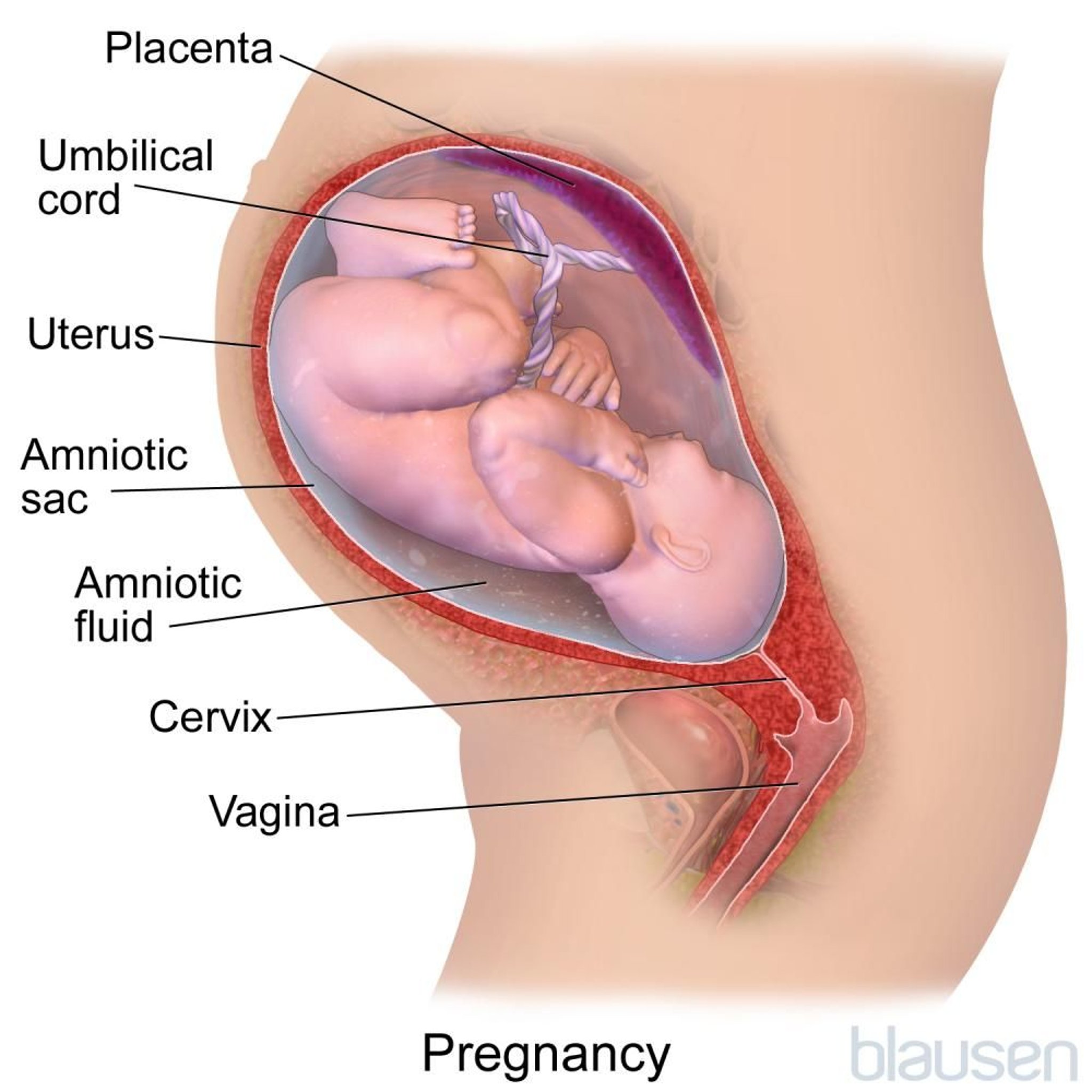
At 8 weeks of pregnancy (6 weeks after fertilization), development of most major organ systems has begun in the embryo. The placenta has also developed and formed tiny finger-like projections (villi) that extend into the wall of the uterus. The villi are part of the circulatory system of the embryo. Blood vessels carry blood from the embryo through the umbilical cord and the placental villi. Then the blood returns to the embryo. Blood vessels from the mother pass next to the placental villi, and maternal blood fills the space around the villi. The blood vessels of the mother and the embryo are separated by a thin membrane. Blood does not run directly from the mother to embryo. Fluid, oxygen, and nutrients pass across the membrane from the mother to the embryo, and carbon dioxide and waste products pass from the embryo to the mother. Cells from the placenta also develop into the amniotic sac. Two layers of membranes form around the embryo: the amnion (inner membrane) and chorion (outer membrane). The amnion and chorion create a sac (amniotic sac) around the embryo. The sac is filled with fluid (amniotic fluid) and the embryo floats in the fluid. The amniotic fluid provides a space in which the embryo can grow freely and helps protect the embryo from injury. The amniotic sac is strong and resilient. |

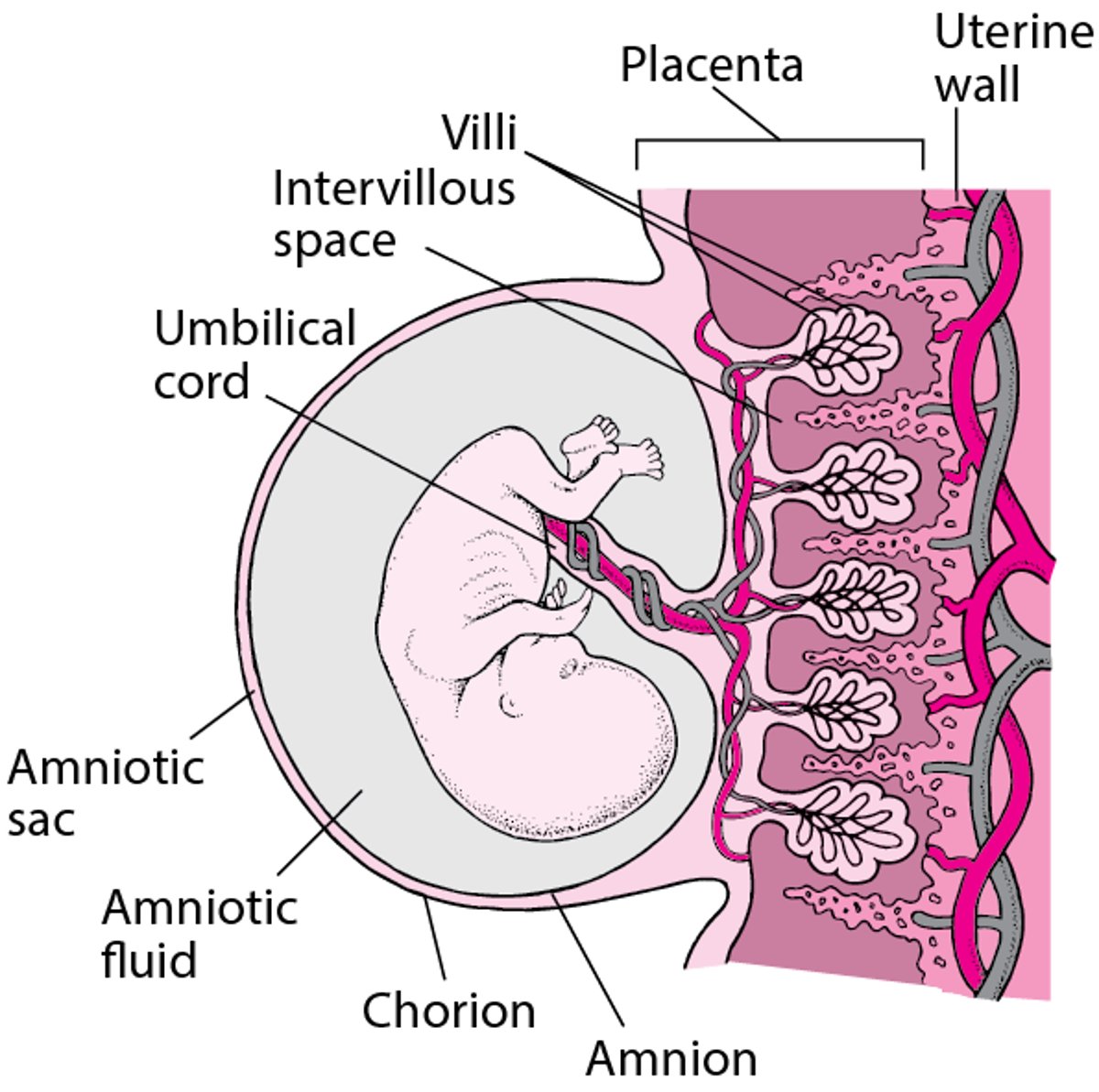
The placenta is fully formed by 18 to 20 weeks but continues to grow throughout pregnancy. At delivery, it weighs about 1 pound.
Development of the Fetus
At the end of the tenth week of pregnancy (8 weeks after fertilization), the fetal stage begins. During this stage, the organs and systems that have already formed grow and develop as follows:
By 12 weeks of pregnancy: The fetus fills the entire uterine cavity. As the pregnancy continues, the uterus enlarges as the fetus grows.
By about 14 weeks: The sex can be identified on ultrasound.
By about 16 to 20 weeks: Typically, the pregnant woman can feel the fetus moving. Women who have been pregnant before typically feel movements about 2 weeks earlier than women who are pregnant for the first time.

The brain accumulates new cells throughout pregnancy and the first year of life after birth. The lungs continue to mature until near the time of delivery.
Pregnancy: Week by Week
The phases of pregnancy are described as trimesters:
| ||
Events | Weeks of Pregnancy* | |
First Trimester | ||
Last menstrual period before fertilization | 1 | 13 Weeks of Pregnancy Image credit: SEBASTIAN KAULITZKI/SCIENCE PHOTO LIBRARY |
Fertilization of an egg by a sperm | 2 | |
Fertilized egg (zygote) divides and develops into a hollow ball of cells called the blastocyst. Blastocyst implants in the wall of uterus. | 3 | |
Outer cells of blastocyst develop into placenta and amniotic sac. Inner cells of blastocyst develop into embryo. | 4 | |
Neural tube, the area that will become the brain and spinal cord, begins to develop. Areas of embryo that will become major organ systems begin to develop. | 5 | |
Heart muscle tissue (myometrium) develops and begins to beat. Buds develop that will become the arms and legs. | 6 | |
Heart chambers (atria and ventricules) form. Blood starts to flow through major blood vessels. | 7 | |
Brain continues to develop. Lungs, digestive system, and kidneys begin to develop. Hands and feet begin to form. | 8 | |
Major organ systems continue to develop. Bones develop and begin to harden. Elbows and toes form. Hair follicles form. | 9 | |
Eyelids and outer ears become more developed. Teeth begin to develop. Intestines rotate. Kidneys begin to function, and the embryo begins to urinate (urine is mostly amniotic fluid). Umbilical cord is fully developed, and blood vessels pass through the cord from the embryo to the placental villi. At the end the 10th week, the fetal stage begins. | 10 | |
Facial features become more developed. All major organ systems have formed but will continue to grow and develop. Liver and spleen begin making red blood cells and platelets. Genitals begin to develop. Nails form on fingers and toes. Fetus starts to open and close hands and mouth. | 11 to 13 | |
Second Trimester | ||
Fetus starts to move. Soft hair (called lanugo) begins to cover the head and body. | 14 to 15 | 27 Weeks of Pregnancy Image credit: SEBASTIAN KAULITZKI/SCIENCE PHOTO LIBRARY |
Fetus moves more vigorously, and the mother starts to be able to feel the movement. Eyes can move, but eyelids stay closed until 27 weeks. Fetus can start hearing sounds. | 16 to 18 | |
A white, cheese-like substance (vernix) starts to cover the fetal skin. Fetus can suck its thumb and can swallow. | 19 to 21 | |
Eyebrows and eyelashes appear. Intestines begin to make meconium (stool). Blood cells begin to be made by the bone marrow. | 22 to 24 | |
The fetus is active, changing positions often. Fetus stores fat. Air sacs begin to form in the lungs. | 25 to 27 | |
Third Trimester | ||
Eyelids open; fetus can blink. Lungs may be developed enough to breathe air. | 28 to 29 | 40 Weeks of Pregnancy Image credit: SEBASTIAN KAULITZKI/SCIENCE PHOTO LIBRARY |
Brain and nervous system become fully developed. Major organ systems are mature, but continue to grow and develop. Fetus starts to practice breathing by breathing in amniotic fluid. Fetus begins to gain weight more rapidly. Fetal head settles into position for birth. | 30 to 36 | |
Labor and delivery | 37 to 42 | |
* Weeks of pregnancy are noted based on the due date, which is usually estimated as 40 weeks after first day of the last menstrual period. The pregnancy actually begins with fertilization of an egg by a sperm, which typically occurs 2 weeks after the last menstrual period. So, development is noted as beginning at 2 weeks of pregnancy. | ||