

Molluscum contagiosum is characterized by clusters of pink, dome-shaped, smooth, waxy, or pearly and umbilicated papules 2 to 5 mm in diameter caused by the molluscum contagiosum virus, a poxvirus. Diagnosis is based on clinical appearance. Treatment is aimed at preventing spread or removing cosmetically unacceptable lesions and can include mechanical methods (eg, curettage, cryosurgery) and topical agents (eg, cantharidin, tretinoin, berdazimer)., tretinoin, berdazimer).
Molluscum contagiosum virus commonly causes a localized chronic infection. Transmission is by direct contact; spread occurs by autoinoculation and via fomites (eg, towels, bath sponges) and bath water. Adults acquire the infection via close skin-to-skin contact with an infected person (eg, sexual contact, wrestling).
Immunocompromised patients (eg, due to HIV/late-stage HIV, corticosteroid use, or chemotherapy) may develop a more widespread infection (1, 2).
Molluscum contagiosum is common among children.
General references
1. Meza-Romero R, Navarrete-Dechent C, Downey C. Molluscum contagiosum: An update and review of new perspectives in etiology, diagnosis, and treatment. Clin Cosmet Investig Dermatol. 12:373–381, 2019. doi: 10.2147/CCID.S187224
2. Edwards S, Boffa MJ, Janier M, et al. 2020 European guideline on the management of genital molluscum contagiosum. J Eur Acad Dermatol Venereol. 35(1):17–26, 2021. doi: 10.1111/jdv.16856
Symptoms and Signs of Molluscum Contagiosum
Molluscum contagiosum can appear anywhere on the skin except the palms and soles. Lesions consist of clusters of pink, dome-shaped, smooth, waxy, or pearly and umbilicated papules, usually 2 to 5 mm in diameter, which occur most commonly on the face, trunk, and extremities in children and on the pubis, penis, or vulva in adults. The latter suggests that the etiology may be sexually transmitted. Lesions may grow to 10 to 15 mm in diameter, especially among patients with HIV infection and other immunodeficiencies.
Lesions are usually not pruritic or painful and may be discovered only coincidentally during a physical examination. However, the lesions can become inflamed and pruritic as the body fights off the virus. Molluscum dermatitis may occur in a subset of patients, which is characterized by eczematous inflammation of contiguous areas surrounding lesions. In children, a Gianotti-Crosti-like syndrome (itchy monomorphic papules on the elbows and knees), may occur in the setting of molluscum contagiosum and usually portends clinical improvement.

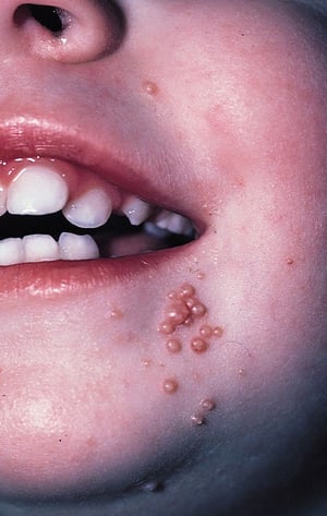
The photo shows lesions of molluscum contagiosum. Lesions are typically 1 to 5 mm, solitary or grouped, firm, painless papules. They are pearly to pink in color, dome shaped, and may be umbilicated.
The photo shows lesions of molluscum contagiosum. Lesions are typically 1 to 5 mm, solitary or grouped, firm, painless
© Springer Science+Business Media

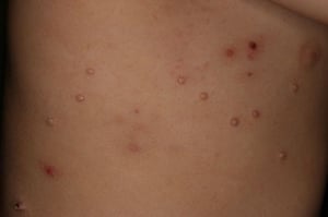
This image shows scattered lesions of molluscum contagiosum on the lateral torso of a toddler.
This image shows scattered lesions of molluscum contagiosum on the lateral torso of a toddler.
Image courtesy of James G.H. Dinulos, MD.

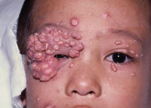
This photo shows very severe lesions on the face of a child with HIV infection. Giant molluscum indicates advanced immunodeficiency.
This photo shows very severe lesions on the face of a child with HIV infection. Giant molluscum indicates advanced immu
© Springer Science+Business Media


This photo shows the firm, dome-shaped, pearly papules with characteristic central umbilication of molluscum contagiosum.
This photo shows the firm, dome-shaped, pearly papules with characteristic central umbilication of molluscum contagiosu
DR HAROUT TANIELIAN/SCIENCE PHOTO LIBRARY
The photo shows lesions of molluscum contagiosum. Lesions are typically 1 to 5 mm, solitary or grouped, firm, painless papules. They are pearly to pink in color, dome shaped, and may be umbilicated.
The photo shows lesions of molluscum contagiosum. Lesions are typically 1 to 5 mm, solitary or grouped, firm, painless
© Springer Science+Business Media
This image shows scattered lesions of molluscum contagiosum on the lateral torso of a toddler.
This image shows scattered lesions of molluscum contagiosum on the lateral torso of a toddler.
Image courtesy of James G.H. Dinulos, MD.
This photo shows very severe lesions on the face of a child with HIV infection. Giant molluscum indicates advanced immunodeficiency.
This photo shows very severe lesions on the face of a child with HIV infection. Giant molluscum indicates advanced immu
© Springer Science+Business Media
This photo shows the firm, dome-shaped, pearly papules with characteristic central umbilication of molluscum contagiosum.
This photo shows the firm, dome-shaped, pearly papules with characteristic central umbilication of molluscum contagiosu
DR HAROUT TANIELIAN/SCIENCE PHOTO LIBRARY
Diagnosis of Molluscum Contagiosum
History and physical examination
Diagnosis of molluscum contagiosum is usually based on clinical appearance. When performed, skin biopsy or smear of expressed material shows characteristic inclusion bodies but is necessary only when diagnosis is uncertain.
Differential diagnosis includes folliculitis, milia, and warts (for lesions < 2 mm) and juvenile xanthogranuloma and Spitz nevus (for lesions > 2 mm).
Treatment of Molluscum Contagiosum
Physical removal: Curettage, cryosurgery, laser therapy, or electrocautery
Topical treatments (eg, berdazimer, trichloroacetic acid, Topical treatments (eg, berdazimer, trichloroacetic acid,cantharidin, tretinoin, tazarotene, podophyllotoxin [podofilox]) , tretinoin, tazarotene, podophyllotoxin [podofilox])
Sometimes intralesional injection or photodynamic therapy
Sometimes combination therapies
Sometimes oral therapy (cimetidine)Sometimes oral therapy (cimetidine)
Most lesions spontaneously regress in 6 months to 2 years, but they can remain for up to 2 to 3 years.
Treatment of molluscum contagiosum is indicated for cosmetic reasons and for prevention of spread. Options include curettage, cryosurgery, laser therapy, electrocautery, trichloroacetic acid (25 to 40% solution), Treatment of molluscum contagiosum is indicated for cosmetic reasons and for prevention of spread. Options include curettage, cryosurgery, laser therapy, electrocautery, trichloroacetic acid (25 to 40% solution),cantharidin, podophyllotoxin (podofilox) in adults, berdazimer, tretinoin, and tazarotene. Some clinicians use salicylic acid, but others consider it too irritating for many body areas where molluscum occurs. Similar concerns exist with the use of potassium hydroxide (KOH). Molluscum lesions within the orbital rim should be removed via gentle destruction by a skilled clinician. Lesions may be gently squeezed with a forceps to remove the central core. , podophyllotoxin (podofilox) in adults, berdazimer, tretinoin, and tazarotene. Some clinicians use salicylic acid, but others consider it too irritating for many body areas where molluscum occurs. Similar concerns exist with the use of potassium hydroxide (KOH). Molluscum lesions within the orbital rim should be removed via gentle destruction by a skilled clinician. Lesions may be gently squeezed with a forceps to remove the central core.
Treatments that cause minimal pain (eg, tretinoin, tazarotene, cantharidin) used first, especially in children. Imiquimod was used in the past due to favorable responses in observational studies; however, it is usually not recommended now due to its lack of proven efficacy in controlled environments and adverse effects including pruritus, crusting, and tenderness over the site of application. Berdazimer 10.3% gel is a topical agent available for use in patients 1 year and older that has demonstrated favorable efficacy (ie, clearing all molluscum lesions in over one-third of patients) and safety in randomized trials () used first, especially in children. Imiquimod was used in the past due to favorable responses in observational studies; however, it is usually not recommended now due to its lack of proven efficacy in controlled environments and adverse effects including pruritus, crusting, and tenderness over the site of application. Berdazimer 10.3% gel is a topical agent available for use in patients 1 year and older that has demonstrated favorable efficacy (ie, clearing all molluscum lesions in over one-third of patients) and safety in randomized trials (1). It is applied at home once daily to the lesion (may not be used over mucous membranes) for up to 12 weeks. This compound increases nitric oxide, which has antimicrobial and antiviral properties. Application-site erythema and pain are the most common adverse effects of berdazimer use; both can range from mild to moderate in severity.). It is applied at home once daily to the lesion (may not be used over mucous membranes) for up to 12 weeks. This compound increases nitric oxide, which has antimicrobial and antiviral properties. Application-site erythema and pain are the most common adverse effects of berdazimer use; both can range from mild to moderate in severity.
Cantharidin is safe and effective but can cause blistering. It is applied in 1 small drop directly to the molluscum lesion. Areas that patients (especially children) may rub are covered with a bandage because contact with the fingers should be avoided. Cantharidin should not be applied to the face or near the eyes because blistering is unpredictable. If cantharidin comes into contact with the cornea, it can cause scarring. Cantharidin should be washed off with soap and water in 6 to 24 hours. Fewer than 15 lesions should be treated in one session because infection may occur after application of cantharidin. Parents should be warned about blistering if their children are prescribed this irritant.
Curettage or cryotherapy (using liquid nitrogen) can be performed 40 to 60 minutes after application of a topical anesthetic such as EMLA (lidocaine/prilocaine) cream or 4% lidocaine cream under an occlusive dressing. EMLA cream must be applied judiciously because it can cause systemic toxicity, especially in children. In adults, curettage is very effective but painful if done without anesthetic. Dermatologists often use combination therapy such as cryotherapy with liquid nitrogen plus Curettage or cryotherapy (using liquid nitrogen) can be performed 40 to 60 minutes after application of a topical anesthetic such as EMLA (lidocaine/prilocaine) cream or 4% lidocaine cream under an occlusive dressing. EMLA cream must be applied judiciously because it can cause systemic toxicity, especially in children. In adults, curettage is very effective but painful if done without anesthetic. Dermatologists often use combination therapy such as cryotherapy with liquid nitrogen pluscantharidin in the office, or a retinoid cream at home. This form of therapy is typically successful, but resolution often takes 1 to 2 months in some patients.
Other treatments include intralesional injection (eg, with Candida antigen or rarely interferon alpha in immunocompromised patients) and photodynamic therapy (2). Antiviral and immunomodulatory medications have been more successful in patients infected with HIV (3, 4). Oral cimetidine is a histamine-receptor antagonist with immunomodulatory properties, has been effective in clearing topical lesions in some pediatric patients. In patients with compromised cellular immunity, or those with severe, recalcitrant molluscum, topical (1% or 3%) or IV cidofovir has also been used. When using cidofovir, care should be taken to avoid renal toxicity (eg, by concurrent administration of probenecid). Before selecting a treatment option, it is helpful to evaluate the strength of evidence backing these therapies, their local availabilities and costs (). Oral cimetidine is a histamine-receptor antagonist with immunomodulatory properties, has been effective in clearing topical lesions in some pediatric patients. In patients with compromised cellular immunity, or those with severe, recalcitrant molluscum, topical (1% or 3%) or IV cidofovir has also been used. When using cidofovir, care should be taken to avoid renal toxicity (eg, by concurrent administration of probenecid). Before selecting a treatment option, it is helpful to evaluate the strength of evidence backing these therapies, their local availabilities and costs (5).
Children should not be excluded from school or day care. However, their lesions should be covered to reduce the risk of spread.
Treatment references
1. Sugarman JL, Hebert A, Browning JC, et al. Berdazimer gel for molluscum contagiosum: An integrated analysis of 3 randomized controlled trials. . Berdazimer gel for molluscum contagiosum: An integrated analysis of 3 randomized controlled trials.J Am Acad Dermatol. 2024;90(2):299-308. doi:10.1016/j.jaad.2023.09.066
2. Wells A, Saikaly SK, Schoch JJ. Intralesional immunotherapy for molluscum contagiosum: A review. Dermatol Ther. 33(6):e14386, 2020. doi: 10.1111/dth.14386
3. Vora RV, Pilani AP, Kota RK. Extensive giant molluscum contagiosum in a HIV positive patient. J Clin Diagn Res. 9(11):WD01-2, 2015. doi: 10.7860/JCDR/2015/15107.6797
4. Dohil M, Prendiville JS. Treatment of molluscum contagiosum with oral cimetidine: clinical experience in 13 patients. . Treatment of molluscum contagiosum with oral cimetidine: clinical experience in 13 patients.Pediatr Dermatol. 1996;13(4):310-312. doi:10.1111/j.1525-1470.1996.tb01247.x
5. Gerlero P, Hernández-Martín Á. Update on the Treatment of molluscum Contagiosum in children. Actualización sobre el tratamiento de moluscos contagiosos en los niños. Actas Dermosifiliogr (Engl Ed). 2018;109(5):408-415. doi:10.1016/j.ad.2018.01.007
Key Points
Molluscum contagiosum, caused by a poxvirus, commonly spreads by direct contact (eg, sexual contact, wrestling), fomites, and bath water.
Lesions tend to be asymptomatic clusters of 2- to 5-mm diameter papules that are pink, dome-shaped, smooth, waxy, or pearly and umbilicated.
Diagnose based on clinical appearance.
Treat for cosmetic reasons or prevention of spread.
Treatments can include destructive methods (eg, curettage, cryosurgery, laser therapy, electrocautery) or topical agents (eg, trichloroacetic acid, Treatments can include destructive methods (eg, curettage, cryosurgery, laser therapy, electrocautery) or topical agents (eg, trichloroacetic acid,cantharidin, tretinoin, tazarotene, podophyllotoxin, berdazimer)., tretinoin, tazarotene, podophyllotoxin, berdazimer).
Drug Information for the Topic


