


 Screener")

Male sexual development and hormonal function depend on a complex feedback circuit involving the hypothalamus-pituitary-gonadal (HPG) axis, which is modulated by the central nervous system. Male sexual dysfunction can be secondary to hypogonadism; trauma; neurovascular, genetic, or other conditions; or medication or recreational drug use.

Physiology
In sexually mature males, the hypothalamus produces gonadotropin-releasing hormone (GnRH), which is released in a pulsatile fashion every 60 to 120 minutes. Its target organ, the anterior pituitary gland, responds to each pulse of GnRH by producing a corresponding pulse of luteinizing hormone (LH) and, to a lesser degree, follicle-stimulating hormone (FSH). If the GnRH pulses do not occur with the proper amplitude, frequency, and diurnal variation, hypogonadism may result (idiopathic hypogonadotropic hypogonadism). Continuous (as opposed to pulsatile) stimulation by GnRH agonists (eg, as a treatment for advanced prostate cancer) actually suppresses pituitary release of LH and FSH, also resulting in hypogonadotropic hypogonadism.
The Leydig cells of the testes respond to LH by producing between 5 and 10 mg of testosterone daily. Testosterone levels are highest in the early morning and lowest during the evening hours; however, in older males, this diurnal pattern may be blunted.
Testosterone is synthesized from cholesterol through several intermediate compounds, including dehydroepiandrosterone (DHEA) and androstenedione. Circulating testosterone is mostly protein bound, approximately 40% avidly bound to sex hormone–binding globulin (SHBG) and 58% loosely bound to albumin. The remaining 2% of circulating testosterone exists as free testosterone. Both free testosterone and the testosterone that is loosely bound to albumin are together referred to as bioavailable testosterone, because these forms can be used by the body. This bioactive component of total testosterone is responsible for male characteristics, libido, bone density, and muscle mass.
In target tissues, approximately 4 to 8% of testosterone is converted to a more potent metabolite, dihydrotestosterone (DHT), by the enzyme 5-alpha-reductase. DHT has important trophic effects in the prostate and mediates androgenetic alopecia. In adults, spermatogenesis requires adequate intratesticular testosterone, but the role of DHT in spermatogenesis is unclear.
Testosterone and DHT have metabolic and other effects, including
Stimulating protein anabolism (increasing muscle mass and bone density)
Stimulating renal erythropoietin production (increasing red blood cell mass)
Stimulating bone marrow stem cells (modulating the immune system)
Causing cutaneous effects (eg, sebum production, hair growth)
Causing neural effects (eg, affecting cognition, increasing libido, and possibly increasing aggression)
Testosterone also undergoes conversion to estradiol by the enzyme aromatase; estradiol mediates most of testosterone's action on organs such as bones and the brain. In males, aromatase is most active in adipose tissue; therefore, patients with obesity are likely to have higher estradiol levels.
Testosterone, DHT, and estradiol provide negative feedback on the hypothalamic-pituitary axis. In males, estradiol is the main inhibitor of LH production, whereas both estradiol and inhibin B, a peptide produced by Sertoli cells of the testes, inhibit production of FSH. In the presence of testosterone, FSH stimulates the Sertoli cells and induces spermatogenesis. In spermatogenesis, each germinal cell (spermatogonium), located adjacent to the Sertoli cells, undergoes differentiation into 16 primary spermatocytes, each of which generates 4 spermatids. Each spermatid matures into a spermatozoon. Spermatogenesis takes 72 to 74 days and yields approximately 100 million new spermatozoa each day. Upon maturation, spermatozoa are released into the rete testis, where they migrate to the epididymis and eventually to the vas deferens. Migration requires an additional 14 days. During ejaculation, spermatozoa are mixed with secretions from the seminal vesicles, prostate, and bulbourethral glands.
Sexual Differentiation, Adrenarche, and Puberty
In the embryo, the presence of a Y chromosome triggers development and growth of the testes, which begin secreting testosterone and a müllerian duct inhibitor by about 7 weeks of gestation. Testosterone virilizes the wolffian duct (which develops into the epididymis, vas deferens, and seminal vesicles). DHT promotes development of the male external genitals. Testosterone levels peak in the second trimester and fall to almost zero by birth. Testosterone production rises briefly during the first 6 months of life, after which testosterone levels remain low until puberty. Müllerian inhibitory factor causes regression of the female genital organs in the fetus.
LH and FSH are elevated at birth but fall to low levels within a few months, remaining low or undetectable throughout the prepubertal years. Through an unknown mechanism, blood levels of the adrenal androgens DHEA and DHEA sulfate begin to increase several years before puberty. Their conversion to testosterone in small amounts initiates pubic and axillary hair growth (adrenarche). Adrenarche can occur as early as 9 or 10 years of age.
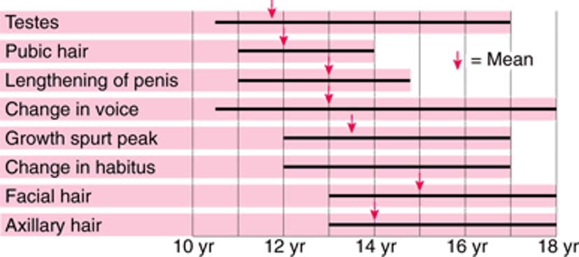
The mechanisms that initiate puberty are unclear, although early in puberty the hypothalamus becomes less sensitive to the inhibitory effects of sex hormones. This desensitization increases secretion of LH and FSH, corresponding to pulsatile GnRH secretion, and stimulating testosterone and sperm production. In boys, the increased testosterone levels cause pubertal changes, the first of which is growth of the testes and scrotum. Later, penile length, muscle mass, and bone density increase; the voice deepens; and pubic and axillary hair becomes denser and thicker (see figure Puberty—when male sexual characteristics develop). (See Physical Growth and Sexual Maturation of Adolescents for more detail.)
Puberty—When Male Sexual Characteristics Develop
Bars indicate normal ranges. No mean is available for change in habitus. |

Effects of Aging
Both hypothalamic secretion of GnRH and the response of Leydig cells to follicle-stimulating hormone FSH and LH diminish with aging. In older males, Leydig cells decrease in number as well. Beginning at about age 30, a man’s serum total testosterone levels decline by 1 to 2% annually (1). Men aged 70 to 80 years tend to have serum testosterone levels that are about one-half to two-thirds of those of men in their 20s. In addition, sex hormone–binding globulin (SHBG) levels increase with aging, causing an even greater decline in bioavailable testosterone. FSH and LH levels tend to be normal or high-normal. These age-related changes are referred to as the andropause, although there are not the abrupt changes in hormone levels (and corresponding symptoms) that occur in menopause. The decline in testosterone may contribute to a combination of symptoms that has been termed androgen deficiency of the aging male (ADAM), which includes
Age-related muscle loss
Increased fat deposition
Osteopenia
Cognitive decline
If men have these symptoms plus low serum testosterone (defined as 2 early morning total testosterone levels both < 300 ng/dL [< 10.41 nmol/L]), they are diagnosed with hypogonadism and are eligible for treatment with supplemental testosterone. One study found that 39% of men ≥ 45 years were hypogonadal (and are eligible for treatment with supplemental testosterone. One study found that 39% of men ≥ 45 years were hypogonadal (2).
Testosterone supplementation in men with low-normal levels (300 to 400 ng/dL [10.41 to 13.88 nmol/L]) of testosterone is controversial. Some experts recommend a trial of testosterone supplementation in older men with symptoms or signs of hypogonadism and whose serum testosterone levels are slightly below the lower limit of normal. No data favor any of the testosterone preparations specifically for use in ADAM.
References
1. Harman SM, Metter EJ, Tobin JD, Pearson J, Blackman MR; Baltimore Longitudinal Study of Aging. Longitudinal effects of aging on serum total and free testosterone levels in healthy men. Baltimore Longitudinal Study of Aging. J Clin Endocrinol Metab 2001;86(2):724-731. doi:10.1210/jcem.86.2.7219
2. Mulligan T, Frick MF, Zuraw QC, Stemhagen A, McWhirter C. Prevalence of hypogonadism in males aged at least 45 years: the HIM study. Int J Clin Pract 2006;60(7):762-769. doi:10.1111/j.1742-1241.2006.00992.x
Drugs Mentioned In This Article


