


 Subclassification System")


 Subclassification System")
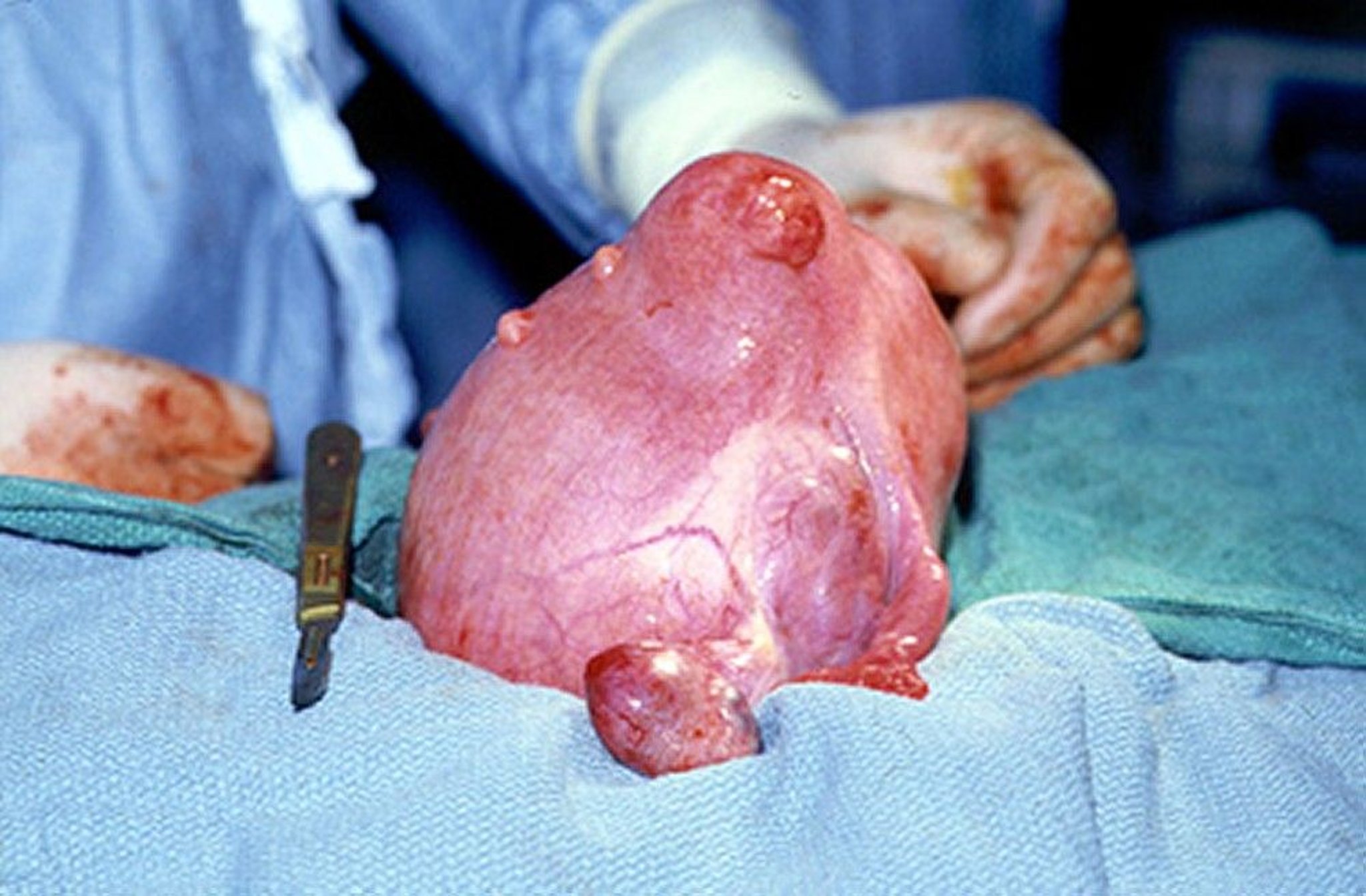
Uterine fibroids (leiomyomas) are smooth muscle tumors that usually arise from the myometrium and are the most common pelvic tumor. Many fibroids are small or asymptomatic.
In the United States, prevalence of uterine fibroids by age 50 is approximately 70% in White women and 80% in Black women (1). Prevalence is increased in women with early menarche, obesity, and hypertension; high parity (3 or more births) is associated with a decreased risk (2).
Although patients are often concerned about cancer in fibroids, sarcomatous change occurs in < 1% of patients (3).
Most patients with fibroids have multiple fibroids. Anatomic locations of fibroids in the uterus are
Subserosal
Intramural
Submucosal
Occasionally, fibroids develop in the broad ligament (intraligamentous), cervix, or, rarely, fallopian tubes. Some fibroids are pedunculated, and others are sessile. Submucosal fibroids may extend into the uterine cavity (intracavitary submucosal fibroids) or prolapse through the uterine cervix (prolapsed fibroid).
The International Federation of Gynecology and Obstetrics (FIGO) classification system for causes of abnormal uterine bleeding (PALM-COEIN system) has a subclassification for location of fibroids and the degree to which they protrude into the endometrial cavity (4).
PALM-COEIN* Uterine Leiomyoma (Fibroid) Subclassification System
Leiomyoma Location Categories | Type | Specific Location in the Uterus |
Submucosal (in contact with the endometrium and/or protruding into the uterine cavity) | 0 | Pedunculated intracavitary |
1 | < 50% intramural | |
2 | ≥ 50% intramural | |
3 | Contacts endometrium, 100% intramural | |
Other: Intramural (within the myometrium); subserosal (in contact with the serosa and/or protruding into the peritoneal cavity); or other locations (eg, cervical, parasitic) | 4 | Intramural |
5 | Subserosal ≥ 50% intramural | |
6 | Subserosal < 50% intramural | |
7 | Subserosal pedunculated | |
8 | Other (specify, eg, cervical, parasitic) | |
Hybrid (in contact with both the endometrium and the serosa) | 2-5† | Image shows one example: Submucosal and subserosal, each with less than half the diameter in the endometrial and peritoneal cavities |
* PALM-COEIN is a mnemonic for the structural causes (PALM) and nonstructural (COEIN) causes of abnormal bleeding (see figure PALM-COEIN Classification System). | ||
† Hybrid leiomyomas are documented as 2 numbers separated by a hyphen. By convention, the first number refers to the relationship with the endometrium and the second number refers to the relationship to the serosa. | ||
Definitions: Endometrium = glandular lining of the uterine cavity (also referred to as the endometrial cavity); myometrium = smooth muscle layer of the uterus, located between the endometrium and serosa; serosa = thin outer lining of the uterus that faces the peritoneal (abdominal) cavity, composed of mesothelium and loose connective tissue. | ||
Adapted from Munro MG, Critchley HOD, Fraser IS; FIGO Menstrual Disorders Committee. The two FIGO systems for normal and abnormal uterine bleeding symptoms and classification of causes of abnormal uterine bleeding in the reproductive years: 2018 revisions [published correction appears in Int J Gynaecol Obstet. 2019 Feb;144(2):237]. Int J Gynaecol Obstet. 2018;143(3):393-408. doi:10.1002/ijgo.12666. | ||


Each fibroid develops from a single smooth muscle cell, making them monoclonal in origin. Because they respond to estrogen, fibroids tend to enlarge during the reproductive years and decrease in size after menopause.
Fibroids may outgrow their blood supply and degenerate. Degeneration is described as hyaline, myxomatous, calcific, cystic, fatty, red (usually only during pregnancy), or necrotic.

(U.S. Navy photograph)
References
1. Baird DD, Dunson DB, Hill MC, et al. High cumulative incidence of uterine leiomyoma in black and white women: ultrasound evidence. Am J Obstet Gynecol 188(1):100-107, 2003. doi:10.1067/mob.2003.99
2. Pavone D, Clemenza S, Sorbi F, et al. Epidemiology and Risk Factors of Uterine Fibroids. Best Pract Res Clin Obstet Gynaecol 46:3-11, 2018. doi:10.1016/j.bpobgyn.2017.09.004
3. Kho KA, Lin K, Hechanova M, Richardson DL. Risk of Occult Uterine Sarcoma in Women Undergoing Hysterectomy for Benign Indications [published correction appears in Obstet Gynecol. 2016 May;127(5):968. doi: 10.1097/AOG.0000000000001427]. Obstet Gynecol. 2016;127(3):468-473. doi:10.1097/AOG.0000000000001242
4. Munro MG, Critchley HOD, Fraser IS; FIGO Menstrual Disorders Committee. The two FIGO systems for normal and abnormal uterine bleeding symptoms and classification of causes of abnormal uterine bleeding in the reproductive years: 2018 revisions [published correction appears in Int J Gynaecol Obstet. 2019 Feb;144(2):237]. Int J Gynaecol Obstet 143(3):393-408, 2018. doi:10.1002/ijgo.12666
Symptoms and Signs of Uterine Fibroids
Many fibroids are asymptomatic; approximately 15 to 30% of patients with fibroids develop severe symptoms (1). Fibroids can cause abnormal uterine bleeding (eg, heavy menstrual bleeding, intermenstrual bleeding). Bleeding can be severe enough to cause anemia.
Bulk symptoms, including chronic pelvic pain or pressure, result from the size or position of fibroids or uterine enlargement due to fibroids (2). Urinary symptoms (eg, urinary frequency or urgency) can result from bladder compression, and intestinal symptoms (eg, constipation) can result from intestinal compression.
Less commonly, fibroids grow and degenerate or torsion of pedunculated fibroids occurs, and severe acute pain can result.
Fibroids may be associated with infertility, especially submucosal fibroids. During pregnancy fibroids are usually asymptomatic, but may cause pain, recurrent spontaneous abortion, premature uterine contractions, placental abruption, or abnormal fetal presentation. Fibroids may also cause postpartum hemorrhage, especially if located in the lower uterine segment (3).
Symptoms and signs references
1. Havryliuk Y, Setton R, Carlow JJ, et al. Symptomatic Fibroid Management: Systematic Review of the Literature. JSLS 21(3):e2017.00041, 2017. doi:10.4293/JSLS.2017.00041
2. Soliman AM, Margolis MK, Castelli-Haley J, Fuldeore MJ, Owens CD, Coyne KS. Impact of uterine fibroid symptoms on health-related quality of life of US women: evidence from a cross-sectional survey. Curr Med Res Opin. 2017;33(11):1971-1978. doi:10.1080/03007995.2017.1372107
3. Qidwai GI, Caughey AB, Jacoby AF. Obstetric outcomes in women with sonographically identified uterine leiomyomata. Obstet Gynecol. 2006;107(2 Pt 1):376-382. doi:10.1097/01.AOG.0000196806.25897.7c
Diagnosis of Uterine Fibroids
Imaging (ultrasound, saline infusion sonography, or MRI)
The diagnosis of uterine fibroids is clinical and is likely if bimanual pelvic examination detects an enlarged, irregular, mobile uterus.
If an enlarged, irregular, mobile uterus is a new finding or if pelvic examination findings have changed (eg, increased uterine size, possible adnexal mass, fixed mass, new finding of tenderness), imaging studies should be performed to evaluate for fibroids or other gynecologic pathology (eg, ovarian masses). Imaging may also be indicated if the patient has new symptoms (eg, bleeding, pain).
Pelvic ultrasound (usually transvaginal) is typically the preferred first-line imaging test (1).
If submucosal fibroids are suspected due to abnormal uterine bleeding or infertility, saline infusion sonography may be performed. In saline infusion sonography, saline is instilled into the uterus, improving visualization of the uterine cavity. Alternatively, hysteroscopy can be used to directly visualize suspected submucosal uterine fibroids and, if needed, to biopsy or resect small fibroids.
MRI is typically performed if an ultrasound or other factors (eg, rapid growth of a presumed fibroid, fixed pelvic mass) suggest a diagnosis of a variant of leiomyomas (eg, smooth muscle tumors of uncertain malignant potential) or a malignant uterine mass (eg, leiomyosarcoma) (2). MRI is also commonly used in patients prior to myomectomy to determine fibroid location.
Patients with postmenopausal bleeding with or without fibroids should be evaluated for uterine cancer.
Diagnosis references
1. Mension E, Carmona F, Vannuccini S, Chapron C. Clinical signs and diagnosis of fibroids from adolescence to menopause. Fertil Steril. 2024;122(1):12-19. doi:10.1016/j.fertnstert.2024.05.003
2. Expert Panel on GYN and OB Imaging, Ascher SM, Wasnik AP, et al. ACR Appropriateness Criteria® Fibroids. J Am Coll Radiol. 2022;19(11S):S319-S328. doi:10.1016/j.jacr.2022.09.019
Treatment of Uterine Fibroids
Hormonal or nonhormonal medications to decrease bleeding (eg, nonsteroidal anti-inflammatory drugs [NSAIDs], tranexamic acid, estrogen-progestin contraceptives, or progestins) Hormonal or nonhormonal medications to decrease bleeding (eg, nonsteroidal anti-inflammatory drugs [NSAIDs], tranexamic acid, estrogen-progestin contraceptives, or progestins)
Myomectomy (to preserve fertility) or hysterectomy
Sometimes other procedures (eg, uterine fibroid embolization)
Treatment options can be classified as medical, procedural, or surgical.
Asymptomatic fibroids do not require treatment. Patients with small and stable size fibroids can be reevaluated periodically (eg, every 12 months).
For symptomatic fibroids, pharmacologic options are typically used first, prior to considering procedural or surgical treatments. Medications are effective in some patients but are often suboptimal. In perimenopausal women with mild symptoms, expectant management is often preferred because symptoms may resolve as fibroids decrease in size after menopause.
Medications to treat fibroids
For control of abnormal uterine bleeding due to fibroids, oral hormonal or nonhormonal medications may be used as first-line therapy. These medications do not decrease fibroid size, and thus do not treat bulk symptoms (eg, pelvic pain and pressure). First-line pharmacologic options include
Estrogen-progestin contraceptives
Progestins (eg, levonorgestrel intrauterine device [IUD])Progestins (eg, levonorgestrel intrauterine device [IUD])
Tranexamic acidTranexamic acid
Nonsteroidal anti-inflammatory drugs (NSAIDs)
For patients who also desire contraception, estrogen-progestin contraceptives or a levonorgestrel IUDlevonorgestrel IUD may be good options.
Exogenous progestins can partially suppress estrogen stimulation of uterine fibroid growth. Oral progestin therapy may be cyclic (10 to 14 days of each menstrual cycle) or continuous (daily); examples of medications and dosages include medroxyprogesterone acetate 5 to 10 mg and megestrol acetate 40 mg. stimulation of uterine fibroid growth. Oral progestin therapy may be cyclic (10 to 14 days of each menstrual cycle) or continuous (daily); examples of medications and dosages include medroxyprogesterone acetate 5 to 10 mg and megestrol acetate 40 mg.
Depot medroxyprogesterone acetate 150 mg IM every 3 months has effects similar to those of continuous oral therapy. Before initiating IM therapy, a trial of oral progestins should be given to determine whether patients can tolerate the adverse effects associated with progestins (eg, weight gain, depression, irregular bleeding). A levonorgestrel-releasing intrauterine device (IUD) is another progestin therapy option. Progestin therapy causes fibroids to grow in some women.Depot medroxyprogesterone acetate 150 mg IM every 3 months has effects similar to those of continuous oral therapy. Before initiating IM therapy, a trial of oral progestins should be given to determine whether patients can tolerate the adverse effects associated with progestins (eg, weight gain, depression, irregular bleeding). A levonorgestrel-releasing intrauterine device (IUD) is another progestin therapy option. Progestin therapy causes fibroids to grow in some women.
Tranexamic acidTranexamic acid (an antifibrinolytic) can reduce uterine bleeding by up to 40% (1).
NSAIDs can be used to treat pain and may slightly decrease bleeding volume (2).
Medications that may be used to reduce fibroid growth in addition to treating abnormal uterine bleeding due to fibroids include
GnRH analogs
Antiprogestins
Selective estrogen receptor modulators (SERMs)
DanazolDanazol
GnRH analogs are either agonists (eg, leuprolide) or antagonists (elagolix and relugolix) that inhibit the hypothalamic-pituitary-ovarian axis and induce hypogonadism, resulting in a decrease in are either agonists (eg, leuprolide) or antagonists (elagolix and relugolix) that inhibit the hypothalamic-pituitary-ovarian axis and induce hypogonadism, resulting in a decrease inestrogen production. In general, these medications should not be used in the long term because rebound growth to pretreatment size within 6 months is common. GnRH analog use is often limited by hypoestrogenic adverse effects such as menopausal symptoms, unfavorable changes in lipid profile, and/or decreased bone density. To prevent bone demineralization when these medications are used long term, clinicians should consider giving patients supplemental estrogen (add-back therapy), such as a low-dose estrogen-progestin combination.
GnRH analogs are used if other medications have not been effective and bleeding is persistent, and the patient is anemic. Alternatively, they are given preoperatively to reduce fibroid and uterine volume, making surgery technically more feasible and reducing blood loss during surgery.
GnRH agonists may be given as follows:
IM or subcutaneously (eg, leuprolide 3.75 mg IM every month, goserelin 3.6 mg subcutaneously every 28 days)IM or subcutaneously (eg, leuprolide 3.75 mg IM every month, goserelin 3.6 mg subcutaneously every 28 days)
As a subdermal pellet
As nasal spray (eg, nafarelin)As nasal spray (eg, nafarelin)
GnRH antagonists are available in oral preparations formulated for low-dose add-back therapy to limit hypoestrogenic adverse effects.
For antiprogestins (eg, mifepristone), the dosage is 5 to 50 mg once a day for 3 to 6 months. This dose is lower than the 200-mg dose used for termination of pregnancy; thus, this dose must be mixed specially by a pharmacist and may not always be available.(eg, mifepristone), the dosage is 5 to 50 mg once a day for 3 to 6 months. This dose is lower than the 200-mg dose used for termination of pregnancy; thus, this dose must be mixed specially by a pharmacist and may not always be available.
SERMs (eg, raloxifene) may help reduce fibroid growth, but whether they can relieve symptoms as well as other medications is unclear.(eg, raloxifene) may help reduce fibroid growth, but whether they can relieve symptoms as well as other medications is unclear.
DanazolDanazol, an androgenic agonist, can suppress fibroid growth but has a high rate of adverse effects (eg, weight gain, acne, hirsutism, edema, hair loss, deepening of the voice, flushing, sweating, vaginal dryness) and is thus rarely used.
Procedures to treat fibroids
Uterine artery embolization is an image-guided treatment option that aims to cause infarction of fibroids throughout the uterus while preserving normal uterine tissue. For this procedure, the uterus is visualized using fluoroscopy, catheters are placed in the femoral artery and advanced into the uterine artery, and then embolic particles are used to occlude blood supply to the fibroids.
After this procedure, women recover more quickly than after hysterectomy or myomectomy, but rates of complications (eg, bleeding, uterine ischemia) and return visits tend to be higher. Treatment failure rates are up to 25% and are higher if bilateral uterine arteries are not embolized (3); in such cases, definitive treatment with hysterectomy is required.
Patients who are considering further childbearing should be counseled that uterine artery embolization may increase certain obstetric outcomes, including spontaneous abortion, cesarean delivery, and postpartum hemorrhage (4).
Magnetic resonance-guided focused ultrasound surgery is a uterine-sparing, percutaneous procedure that uses high-intensity ultrasound waves to ablate fibroids. Data are limited about the safety of pregnancy after this procedure, and further investigation is needed (5).
Surgery for fibroids
Surgery is usually reserved for women with one or more of the following characteristics:
A rapidly enlarging pelvic mass
Recurrent uterine bleeding refractory to medications
Severe or persistent pain or pressure (eg, that requires pain medications for control or that is intolerable to the patient)
A large uterus that has a mass effect in the abdomen, causing urinary or intestinal symptoms or compressing other organs and causing dysfunction (eg, hydronephrosis, urinary frequency, dyspareunia)
Infertility (if submucosal fibroids may be interfering with conception)
Recurrent spontaneous abortion (if pregnancy is desired)
Other factors favoring surgery are a patient's desire for definitive treatment.
For patients with severe bleeding, gonadotropin-releasing hormone (GnRH) agonists may be given before surgery to shrink fibroid tissues; these medications often stop menses and allow blood counts to increase.
Radiofrequency fibroid ablation uses real-time ultrasound to identify the fibroids and apply radiofrequency energy from a handpiece using a laparoscopic or transcervical approach.
Myomectomy can be performed open, laparoscopically or hysteroscopically (using an instrument with a wide-angle telescope and electrical wire loop for excision), with or without robotic techniques.
Hysterectomy can also be done laparoscopically, vaginally, or by laparotomy.
Most indications for myomectomy and hysterectomy are similar, and patients should be counseled about risks and benefits of each procedure.
If women desire future pregnancy or uterine conservation, myomectomy is performed (6). Multiple myomectomy can be more technically difficult than hysterectomy. Multiple myomectomy often involves increased bleeding, postoperative pain, and adhesions, and may increase risk of uterine rupture during subsequent pregnancies.
Factors that favor hysterectomy include
The patient does not desire future childbearing.
Hysterectomy is a definitive treatment. After myomectomy, new fibroids may begin to grow again, and about 25% of women who have a myomectomy have a hysterectomy about 4 to 8 years later.
The patient has other abnormalities that make a more complex surgery like myomectomy more complicated (eg, extensive adhesions, endometriosis).
Hysterectomy would treat or decrease the risk of another disorder (eg, cervical intraepithelial neoplasia, endometrial hyperplasia, endometriosis, ovarian cancer in women with a BRCA mutation, Lynch syndrome).
If hysterectomy or myomectomy is done laparoscopically, techniques must be used to remove the fibroid tissue through the small laparoscopic incisions or vagina. Morcellation is a term that describes cutting fibroids or uterine tissue into small pieces; this may be done with a scalpel or an electromechanical device. Morcellation should not be used in patients with suspected uterine cancer or significant risk factors, particularly for uterine sarcoma. Prior to surgery for presumed fibroids, patients should be evaluated for uterine cancer (7). Women who have surgery for presumed uterine fibroids may have an unsuspected, undiagnosed sarcoma or other uterine cancer; estimated incidence varies from 1 in 300 to < 1 in 10,000 surgeries (8). If intraperitoneal morcellation is done, malignant cells may be disseminated throughout the peritoneum. Surgeons may use methods to prevent dissemination of tissue during morcellation, including extraperitoneal morcellation (tissue is grasped and pulled through the incision or vagina) or use of an intraabdominal bag to contain tissue.
Choice of treatment
Treatment of uterine fibroids should be individualized, but the following factors can help with the decision:
Asymptomatic fibroids: No treatment, continue to follow patient
Symptomatic fibroids, particularly if future pregnancy is desired: Oral medications or myomectomy
Severe symptoms when other treatments were ineffective, particularly if pregnancy is not desired: Uterine artery embolization or hysterectomy (high-intensity focused sonography is used in some countries)
Postmenopausal women: Patients with postmenopausal bleeding should be evaluated for uterine cancer. If results are benign or pressure symptoms are the main issues, a trial of expectant management is reasonable (because symptoms tend to remit as fibroids decrease in size after menopause)
Treatment references
1. Lukes AS, Moore KA, Muse KN, et al. Tranexamic acid treatment for heavy menstrual bleeding: a randomized controlled trial. . Tranexamic acid treatment for heavy menstrual bleeding: a randomized controlled trial.Obstet Gynecol. 2010;116(4):865-875. doi:10.1097/AOG.0b013e3181f20177
2. Bofill Rodriguez M, Dias S, Jordan V, et al. Interventions for heavy menstrual bleeding; overview of Cochrane reviews and network meta-analysis. Cochrane Database Syst Rev. 2022;5(5):CD013180. Published 2022 May 31. doi:10.1002/14651858.CD013180.pub2
3. Spies JB, Bruno J, Czeyda-Pommersheim F, Magee ST, Ascher SA, Jha RC. Long-term outcome of uterine artery embolization of leiomyomata. Obstet Gynecol. 2005;106(5 Pt 1):933-939. doi:10.1097/01.AOG.0000182582.64088.84
4. Homer H, Saridogan E. Uterine artery embolization for fibroids is associated with an increased risk of miscarriage. Fertil Steril. 2010;94(1):324-330. doi:10.1016/j.fertnstert.2009.02.069
5. Zou M, Chen L, Wu C, Hu C, Xiong Y. Pregnancy outcomes in patients with uterine fibroids treated with ultrasound-guided high-intensity focused ultrasound. BJOG. 2017;124 Suppl 3:30-35. doi:10.1111/1471-0528.14742
6. Practice Committee of the American Society for Reproductive Medicine. Electronic address: ASRM@asrm.org; Practice Committee of the American Society for Reproductive Medicine. Removal of myomas in asymptomatic patients to improve fertility and/or reduce miscarriage rate: a guideline. Fertil Steril. 2017;108(3):416-425. doi:10.1016/j.fertnstert.2017.06.034
7. American College of Obstetricians and Gynecologists’ Committee on Gynecologic Practice. Uterine Morcellation for Presumed Leiomyomas: ACOG Committee Opinion, Number 822 [published correction appears in Obstet Gynecol. 2021 Aug 1;138(2):313]. Obstet Gynecol 137(3):e63-e74, 2021. doi:10.1097/AOG.0000000000004291
8. Hartmann KE, Fonnesbeck C, Surawicz T, et al. Management of Uterine Fibroids [Internet]. Rockville (MD): Agency for Healthcare Research and Quality (US); 2017 Dec. (Comparative Effectiveness Review, No. 195.)
Key Points
In the United States, prevalence of uterine fibroids by age 50 is approximately 70% in White women and 80% in Black women.
If necessary, confirm the diagnosis with imaging, usually ultrasound (sometimes with saline infusion sonography) or MRI.
For temporary relief of minor symptoms, consider medications (eg, estrogen-progestin contraceptives, tranexamic acid, progestins, or GnRH analogs).For temporary relief of minor symptoms, consider medications (eg, estrogen-progestin contraceptives, tranexamic acid, progestins, or GnRH analogs).
For more lasting relief, consider surgery (eg, myomectomy or other uterus-conserving procedures, particularly if fertility may be desired; hysterectomy for definitive therapy).
Drugs Mentioned In This Article


