- How To Do an Infraorbital Nerve Block, Percutaneous
- How To Do an Ophthalmic Nerve Block
- How To Do an Ulnar Nerve Block
- How To Do a Median Nerve Block
- How To Do a Radial Nerve Block
- How To Do a Digital Nerve Block
- How To Do an Ultrasound-Guided Peripheral Nerve Block
- How To Do Local Wound Infiltration
- How To Do Procedural Sedation and Analgesia
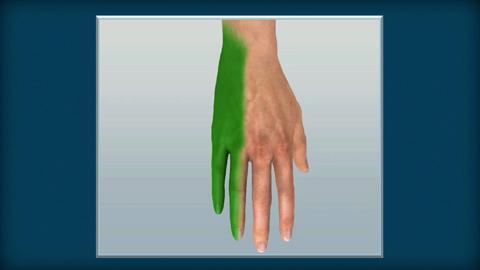
An ulnar nerve block anesthetizes both the volar and dorsal surfaces of the hypothenar half of the hand (from the fifth digit through the ulnar half of the fourth digit).
Topic Resources
Ulnar nerve block can be done using anatomic landmarks or ultrasound guidance. Ultrasound guidance increases the likelihood of successful peripheral nerve blockade and reduces the risk of complications but requires equipment and trained personnel.
(See also Local anesthesia for laceration treatment.)
Indications for Ulnar Nerve Block
Laceration or other surgically treated lesion of the volar or dorsal surface of the ulnar (medial) half of the hand, including the fifth digit and ulnar half of the fourth digit*
Ring removal from the fifth digit or fourth digit
A nerve block has advantages over local anesthetic infiltration because it can cause less pain (eg, in palmar skin repair) and does not distort tissue
* To anesthetize the entire palm, also do a median nerve block.
Contraindications to Ulnar Nerve Block
Absolute contraindications
History of allergy to the anesthetic agent
Absence of anatomic landmarks needed to guide needle insertion (eg, due to trauma)
Relative contraindications
Infection in the path of needle insertion: Use procedural sedation or a different means of anesthesia.
Coagulopathy*: When feasible, correct prior to procedure, or use a different means of analgesia.
* Anticoagulant medications (eg, for atrial fibrillation) increase the risk of bleeding with nerve blocks, but this must be balanced against the increased risk of thrombosis (eg, stroke) if anticoagulation is reversed. Discuss any contemplated reversal with the clinician managing the patient's anticoagulation and then with the patient.
Complications of Ulnar Nerve Block
Adverse reaction to the anesthetic (see Local anesthesia for laceration treatment)
Toxicity due to anesthetic overdose (eg, seizure, cardiac arrhythmias) or sympathomimetic effects due to epinephrine (if using an anesthetic-epinephrine mixture)Toxicity due to anesthetic overdose (eg, seizure, cardiac arrhythmias) or sympathomimetic effects due to epinephrine (if using an anesthetic-epinephrine mixture)
Intravascular injection of anesthetic or epinephrine Intravascular injection of anesthetic or epinephrine
Hematoma
Neuritis
Spread of infection, by passing the needle through an infected area
Most complications result from inaccurate needle placement.
Equipment for Ulnar Nerve Block
Gloves (sterile gloves are not required)
Personal protective equipment as indicated (eg, face mask, safety glasses or face shield, cap and gown)
Antiseptic solution (eg, chlorhexidine, povidone-iodine, alcohol)Antiseptic solution (eg, chlorhexidine, povidone-iodine, alcohol)
Injectable local anesthetic* such as lidocaine 2% with epinephrine† 1:100,000, or for longer-duration anesthesia, bupivacaine 0.5% with epinephrine† 1:200,000 Injectable local anesthetic* such as lidocaine 2% with epinephrine† 1:100,000, or for longer-duration anesthesia, bupivacaine 0.5% with epinephrine† 1:200,000
Syringe (eg, 10 mL) and needle (eg, 25 or 27 gauge, 3.5 cm long)
For ultrasonography: Ultrasound machine with high frequency (eg, 7.5 MHz or higher) linear array probe (transducer); probe cover (eg, transparent sterile dressing, single-use probe cover); sterile, water-based lubricant, single-use packet (preferred over multi-use bottle of ultrasound gel)
* Local anesthetics are discussed in Local anesthesia for laceration treatment.
† Maximum dose of local anesthetics: Lidocaine without epinephrine, 5 mg/kg; lidocaine with epinephrine, 7 mg/kg; bupivacaine, 1.5 mg/kg. NOTE: A 1% solution (of any substance) represents 10 mg/mL (1 g/100 mL). Epinephrine causes vasoconstriction, which prolongs the anesthetic effect. Patients with cardiac disease should receive only limited amounts of epinephrine (maximum 3.5 mL of solution containing 1:100,000 epinephrine); alternatively, use local anesthetic without epinephrine. † Maximum dose of local anesthetics: Lidocaine without epinephrine, 5 mg/kg; lidocaine with epinephrine, 7 mg/kg; bupivacaine, 1.5 mg/kg. NOTE: A 1% solution (of any substance) represents 10 mg/mL (1 g/100 mL). Epinephrine causes vasoconstriction, which prolongs the anesthetic effect. Patients with cardiac disease should receive only limited amounts of epinephrine (maximum 3.5 mL of solution containing 1:100,000 epinephrine); alternatively, use local anesthetic without epinephrine.
Additional Considerations for Ulnar Nerve Block
Document any preexisting nerve deficit in the medical record before doing a nerve block.
Stop the nerve block procedure if you are unsure where the needle is or if the patient is uncooperative. Consider procedural sedation for patients who are unable to cooperate.
Relevant Anatomy for Ulnar Nerve Block
The ulnar nerve lies along the ulnar aspect of the wrist, medial (ulnar) to the ulnar artery.
The artery and the nerve are deep to the flexor carpi ulnaris tendon, which inserts on the pisiform.
The dorsal cutaneous branches of the nerve wrap around the ulna to innervate the dorsum of the ulnar side of the hand.
Positioning for Ulnar Nerve Block
Position the patient with the arm resting with the palm facing up.
Step-by-Step Description of Ulnar Nerve Block
Check sensation and motor function of the ulnar nerve.
Wear gloves and use appropriate personal protective equipment.
Locate the flexor carpi ulnaris tendon by palpating just proximal to the pisiform bone as the patient flexes the wrist against resistance.
The needle-entry site: The needle will be inserted horizontally into the ulnar side of the wrist at the proximal flexor crease, under the flexor carpi ulnaris tendon and distal to the ulnar styloid.
Cleanse the site with antiseptic solution.
Place a skin wheal (shallow intradermal injection) of anesthetic, if one is being used, at the needle-entry site.
Insert the needle horizontally, under the flexor carpi ulnaris tendon, and advance it about 1 to 1.5 cm laterally (radially) toward the ulnar nerve. If paresthesia occurs during insertion, withdraw the needle 1 to 2 mm.
Aspirate to exclude intravascular placement and then slowly (ie, over 30 to 60 seconds) inject 3 to 5 mL of anesthetic.
Next, anesthetize the dorsal branches of the ulnar nerve: Withdraw the needle, then reinsert it dorsally and slowly infiltrate an additional 5 to 6 mL of anesthetic subcutaneously from the lateral border of the flexor carpi ulnaris tendon to the dorsal midline.
Allow about 5 to 10 minutes for the anesthetic to take effect.
Ulnar nerve block, ultrasound-guided
Set the ultrasound machine to 2-D mode or B mode. Adjust the screen settings and probe position if needed to attain an accurate left-right orientation. This almost always means orienting the side-mark on the probe to the operator's left side (corresponding to the left-sided marker dot/symbol on the ultrasound screen).
Cleanse the sides and volar surface of the wrist with antiseptic solution.
Cover the probe tip with a layer of gel, then cover the tip with a sterile transparent dressing tightly (to eliminate air bubbles underneath). Apply sterile lubricant to the covered tip.
Place the probe tip transversely (short-axis, cross-sectional view) on the proximal wrist crease.
Adjust the gain on the console so that the blood vessels are hypoechoic (appear black on the ultrasound screen) and the surrounding tissues are gray. Nerves appear as an echogenic (white), honeycombed, triangular shape, often adjacent to an artery.
Adjust maximum depth to roughly twice the distance from the surface to the ulnar artery.
Slide the probe transversely as needed to center the artery on the ultrasound screen. Identify the ulnar nerve medially adjacent to the artery.
Slowly slide the probe up the wrist to more clearly see the nerve and artery, with some space between them. Move the probe proximal to the distal third of the forearm to ensure placement of the block proximal to the superficial cutaneous nerve branches. Do not move the probe from this spot.
Insert the needle and slightly tilt/rotate the probe to view the needle on the ultrasound screen (an in-plane, longitudinal image).
Maintaining the entire longitudinal needle image on the screen, advance the needle until the tip lies close to the nerve.
Inject a small test dose of anesthetic (about 0.25 mL) to see whether it spreads around the nerve. If not, move the needle closer to the nerve and inject another test dose.
When the needle tip is properly positioned, inject 1 to 2 mL of anesthetic solution to further surround the nerve. If necessary, reposition the needle tip and inject more small amounts; however, the donut sign—nerve completely surrounded by anesthetic—is not required.
Aftercare for Ulnar Nerve Block
Ensure hemostasis at the injection site.
Instruct patient regarding anticipated time to anesthesia resolution.
Warnings and Common Errors for Ulnar Nerve Block
To minimize the risk of needle breakage, do not bend a needle, insert it to its full depth (ie, to the hub), or attempt to change direction of the needle while it is inserted.
To help prevent nerve injury or intraneural injection, instruct patients to report paresthesias or pain during the procedure.
To help prevent intravascular injections, aspirate before injecting.
Always maintain ultrasound visualization of the needle tip during insertion.
If using ultrasound, always maintain ultrasound visualization of the needle tip during insertion.
Tricks and Tips for Ulnar Nerve Block
Minimize the pain of injection by injecting slowly (eg, 30 to 60 seconds), warming the anesthetic solution to body temperature, and buffering the anesthetic with sodium bicarbonate.by injecting slowly (eg, 30 to 60 seconds), warming the anesthetic solution to body temperature, and buffering the anesthetic with sodium bicarbonate.
Some clinicians prefer to use an insulin syringe and volar approach for a landmark-based ulnar nerve block, inserting the needle just lateral (radial) to the flexor carpi ulnaris tendon between the proximal and distal flexor wrist creases; however, this technique may increase the risk of ulnar arterial injection.Some clinicians prefer to use an insulin syringe and volar approach for a landmark-based ulnar nerve block, inserting the needle just lateral (radial) to the flexor carpi ulnaris tendon between the proximal and distal flexor wrist creases; however, this technique may increase the risk of ulnar arterial injection.

