Topic Resources
Pain caused by kidney disorders usually is felt in the side (flank) or small of the back. Occasionally, the pain extends to the center of the abdomen. Usually pain occurs because the kidney’s outer covering (renal capsule) is stretched by a disorder that causes rapid swelling of the kidney or because a stone has entered one of the ureters (tubes connecting the kidney to the bladder). Severe kidney pain is often accompanied by nausea and vomiting.
(See Overview of Urinary Tract Symptoms.)
Causes of Flank Pain
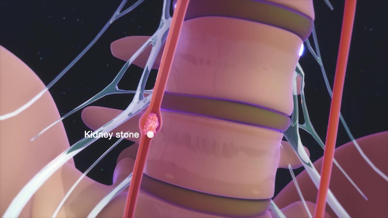
A kidney stone causes excruciating pain when it enters a ureter. The ureter contracts in response to the stone, causing severe, crampy pain (renal or ureteral colic) in the flank or lower back that often extends to the groin or, in men, to a testis. The pain typically comes in waves. A wave may last 20 to 60 minutes and then stop. The pain stops without resuming again when the ureter relaxes or the stone passes into the bladder.
A kidney infection (pyelonephritis) causes swelling of the kidney tissue, which stretches the renal capsule, causing steady, aching pain. Kidney tumors do not usually cause pain until they have become very large.
Other disorders that cause pain in the flank include sudden blockage of blood flow to a kidney or the intestine, ruptured and occasionally unruptured abdominal aortic aneurysms, problems with the spine or spinal nerves, musculoskeletal injuries, and tumors that involve the back of the abdomen (retroperitoneum).
Evaluation of Flank Pain
After noting symptoms, the doctor examines the person and usually does a urinalysis to check for red blood cells or excess white blood cells. White blood cells in the urine suggest an infection. If an infection is suspected, a urine culture is usually done. A person with very severe, cramping pain and blood in the urine is very likely to have a kidney stone. A person with milder, steady pain, tenderness when the doctor taps over 1 kidney, fever, and excess white blood cells in the urine is likely to have a kidney infection.
If a kidney stone is suspected, the doctor often does computed tomography (CT) or ultrasonography to determine whether a stone is the cause, the size and location of the stone, and whether it significantly blocks urine flow. An intravenous contrast agent is not used for the CT scan. If the doctor is not sure of the cause of pain, often CT that uses an intravenous contrast agent or another imaging test is done.
Treatment of Flank Pain
The underlying disorder is treated. Mild pain can be relieved by taking acetaminophen or nonsteroidal anti-inflammatory drugs (NSAIDs). Pain from kidney stones may be severe and may require use of intravenous or oral opioids.The underlying disorder is treated. Mild pain can be relieved by taking acetaminophen or nonsteroidal anti-inflammatory drugs (NSAIDs). Pain from kidney stones may be severe and may require use of intravenous or oral opioids.
Drugs Mentioned In This Article


