- Introduction to Diagnosis of Heart and Blood Vessel Disorders
- Medical History and Physical Examination for Heart and Blood Vessel Disorders
- Electrocardiography
- Continuous Ambulatory Electrocardiography
- Echocardiography and Other Ultrasound Procedures
- X-Rays of the Chest
- Computed Tomography (CT) of the Heart
- Positron Emission Tomography (PET) of the Heart
- Magnetic Resonance Imaging (MRI) of the Heart
- Radionuclide Imaging of the Heart
- Tilt Table Testing
- Electrophysiologic Testing
- Stress Testing
- Central Venous Catheterization
- Pulmonary Artery Catheterization
- Cardiac Catheterization and Coronary Angiography
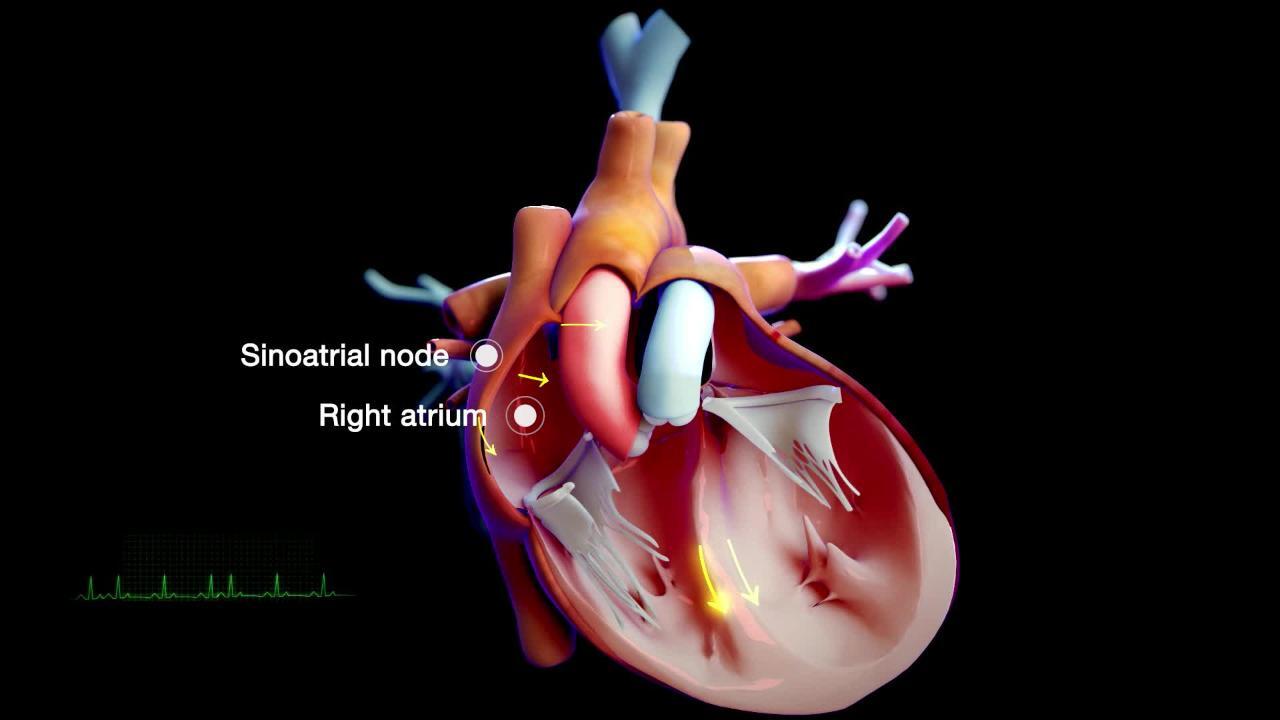
Electrophysiologic testing measures the heart's electrical activity through wire electrodes inserted into all 4 heart chambers.
Topic Resources
Electrophysiologic testing is used to evaluate serious abnormalities in heart rhythm or electrical conduction (see Overview of Abnormal Heart Rhythms).
In people in whom an arrhythmia is already documented or is highly suspected, electrophysiologic testing is used to define precisely the abnormal heart rhythm, determine its cause, and guide treatment. A doctor may intentionally provoke an abnormal heart rhythm during testing to find out whether a particular medication can stop the disturbance or whether an operation will help by eliminating abnormal electrical connections within the heart. If necessary, a doctor can quickly restore a normal rhythm with a brief electrical shock to the heart (cardioversion). Although electrophysiologic testing is an invasive procedure and an anesthetic is required, the procedure is safe. The risk of death is 1 in 5,000. This procedure usually takes 1 to 2 hours.
How electrophysiologic testing is done
Testing is done in the hospital. After injecting a local anesthetic, a doctor inserts a catheter with tiny electrodes at its tip through a needle puncture of a vein in the groin, arm, or neck. The catheter is threaded through the major blood vessels into the heart chambers, usually using fluoroscopy (a continuous x-ray procedure) for guidance. The catheter is used to record the electrocardiogram (ECG) from within the heart and to identify the precise location of the electrical conduction pathways.
Radiofrequency ablation is sometimes done during the procedure and uses heat generated by the radio waves to destroy any abnormal electrical connections in the heart and prevent the person from having further arrhythmias without the need for ongoing medication therapy.
Cryoablation is similar to radiofrequency ablation but uses freezing rather than heat to destroy any abnormal electrical connections.


