


")
")
")

The cause of seborrheic keratosis is unknown, but genetic mutations have been identified in certain types. The lesions commonly occur in middle age and later and most often appear on the trunk or temples. In patients with dark skin, multiple 1- to 3-mm lesions can occur on the cheekbones; this condition is termed dermatosis papulosa nigra.
Seborrheic keratoses vary in size and grow slowly. They may be round or oval and flesh-colored, brown, or black. They usually have a stuck-on appearance and may have a verrucous, velvety, waxy, scaling, or crusted surface.
Seborrheic keratoses that are large, multiple, and/or rapidly developing can be a cutaneous paraneoplastic syndrome (Leser-Trélat sign) in patients who have certain cancers (eg, lymphoma, gastrointestinal cancer).

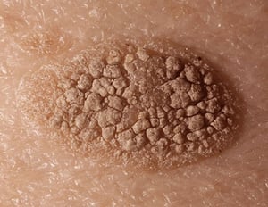
Seborrheic keratoses are benign pigmented lesions. Cause is unknown. They tend to develop in older adults and have a stuck-on appearance with a verrucous, velvety, waxy, scaly, or crusted surface.
Image provided by Thomas Habif, MD.

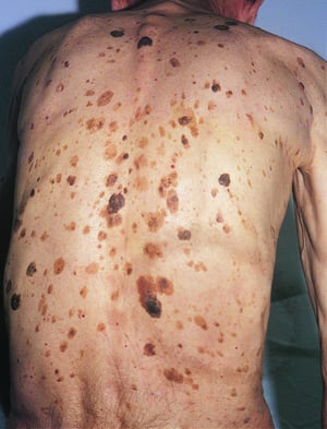
This photo shows seborrheic keratoses (hyperpigmented lesions with a stuck-on appearance) on a patient's back.
DermPics/SCIENCE PHOTO LIBRARY

This photo shows multiple, small seborrheic keratoses on the cheekbones and forehead of a person with dark skin.
DermPics/SCIENCE PHOTO LIBRARY


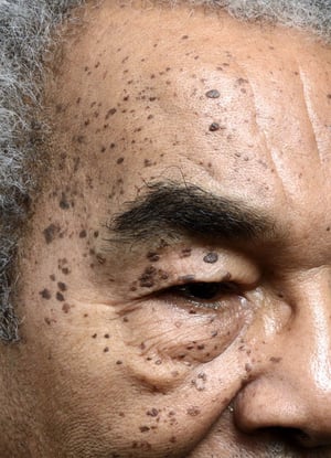
This photo depicts several seborrheic keratoses on the cheekbones and infraorbital region in a patient with dark skin.
Photo courtesy of Karen McKoy, MD.

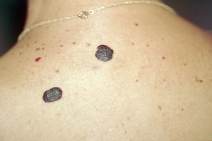
Leser-Trélat sign is the rapid onset of numerous seborrheic keratoses (benign, often pigmented skin lesions with a "stuck-on" appearance).
© Springer Science+Business Media
Seborrheic keratoses are benign pigmented lesions. Cause is unknown. They tend to develop in older adults and have a stuck-on appearance with a verrucous, velvety, waxy, scaly, or crusted surface.
Image provided by Thomas Habif, MD.
This photo shows seborrheic keratoses (hyperpigmented lesions with a stuck-on appearance) on a patient's back.
DermPics/SCIENCE PHOTO LIBRARY
This photo shows multiple, small seborrheic keratoses on the cheekbones and forehead of a person with dark skin.
DermPics/SCIENCE PHOTO LIBRARY
This photo depicts several seborrheic keratoses on the cheekbones and infraorbital region in a patient with dark skin.
Photo courtesy of Karen McKoy, MD.
Leser-Trélat sign is the rapid onset of numerous seborrheic keratoses (benign, often pigmented skin lesions with a "stuck-on" appearance).
© Springer Science+Business Media
Diagnosis of Seborrheic Keratoses
History and physical examination alone
Diagnosis of seborrheic keratosis is clinical. Dermoscopy is very helpful in making a definitive diagnosis of seborrheic keratosis.
Treatment of Seborrheic Keratoses
Removal only if bothersome
Lesions are not premalignant and do not require treatment unless they are irritated, itchy, or cosmetically bothersome to the patient.
Lesions may be removed with little or no scarring by cryotherapy (which can cause hypopigmentation) or by electrodesiccation and curettage after local injection of lidocaine.Lesions may be removed with little or no scarring by cryotherapy (which can cause hypopigmentation) or by electrodesiccation and curettage after local injection of lidocaine.
Drugs Mentioned In This Article


