


Retronychia is one of the most common nail disorders. Acute symptoms include pain, inflammation of the proximal nail fold, xanthonychia, onycholysis, and the presence of granulation tissue. Chronic retronychia is characterized by onycholysis and layers of stacked nail plates. Diagnosis is primarily clinical, but can be confirmed by ultrasound. Treatment varies by stage and may include changes in footwear, nail softening agents, debridement, topical or intralesional glucocorticoids, or camouflage.

")

")
In retronychia the proximal nail plate becomes embedded into the proximal nail fold with loss of the normal alignment of the nail matrix and nail plate. Growth of new nail pushes the old nail upwards, leading to stacking of multiple generations of nail plates.
Retronychia most often affects the hallux (great toenail). It is caused by repetitive microtrauma to the nail, most often by wearing tight shoes or taking part in activities that perpetuate toenail trauma.
Symptoms and Signs of Retronychia
In the acute stage, retronychia is painful and manifests with inflammation of the proximal nail fold, yellow or white discoloration of the nail plate (xanthonychia), and granulation tissue formation. Clinical features that help differentiate retronychia from paronychia include the presence of xanthonychia and swelling of the proximal nail folds rather than the lateral nail folds because the lateral nail folds are affected much less often in retronychia than in paronychia.


Image courtesy of Chris G. Adigun, MD.

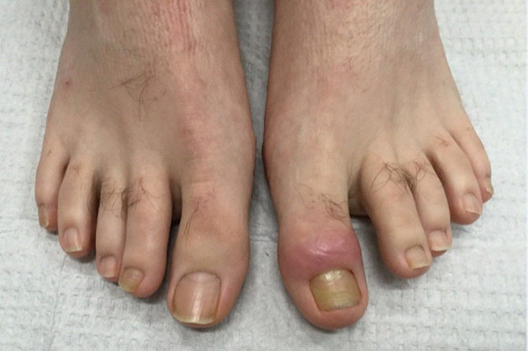
Image courtesy of Chris G. Adigun, MD.
In the chronic stage, the inflammation resolves, but the nail plate becomes thickened and can separate from the nail bed (onycholysis). Layers of nail plates can become stacked, and the cuticle is missing.


Image courtesy of Chris G. Adigun, MD.

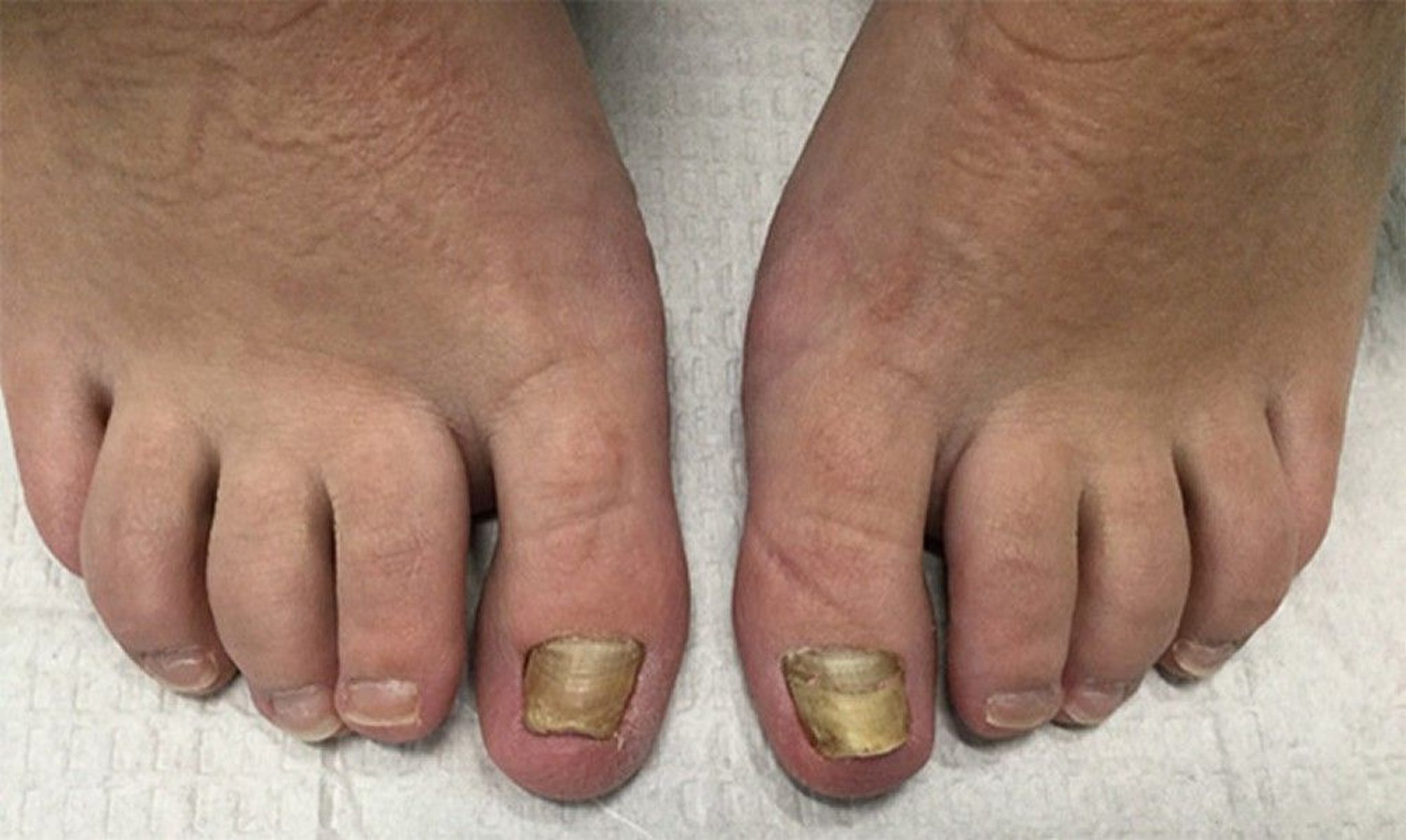
Image courtesy of Chris G. Adigun, MD.
(See also Acute Paronychia and Chronic Paronychia.)
Diagnosis of Retronychia
Primarily physical examination
Periodic acid-Schiff (PAS) stain
Culture
Sometimes ultrasound
The diagnosis of retronychia is primarily based on physical examination. Periodic acid-Schiff (PAS) stain or culture of the nail plate is often negative. Although the nail plate itself may have a positive PAS result if the nail plate is analyzed after removal, the onychomycosis is often not the primary problem but rather secondary to the trauma to the nail that caused compromise and inflammation. The diagnosis includes noticing the layered nail plate, absent cuticle (in chronic retronychia), and absent subungual debris. Ultrasound may be helpful but requires specialized expertise.
Treatment of Retronychia
Glucocorticoids, softening agents, and debridement.
In the acute stage, treatment options include topical and intralesional glucocorticoids (1). In the chronic stage, treatment is softening agents and debridement. Nail avulsion can be performed, but recurrence is common due to underlying mechanical and podiatric issues.
Avoidance of microtrauma, for example, by wearing shoes with more space around the toes to relieve pressure on the affected toenail, is important. Camouflage (using nail polish or lacquers) is an important option to offer patients and can improve quality of life (2).
Treatment references
1. Costa TPE, João AL, Lencastre A: Retronychia: A paradigm shift? Skin Appendage Disord 6(5):268–271, 2020. doi: 10.1159/000509370
2. Curtis KL, Ricardo JW, Shaikh B, et al. Prosthetic resin nail improves quality of life in patients with retronychia in a single-center clinical trial. J Am Acad Dermatol. 2024;90(6):1277-1278. doi:10.1016/j.jaad.2024.01.073


