


Ocular mucous membrane pemphigoid is an autoimmune disease in which binding of anticonjunctival basement membrane antibodies results in conjunctival inflammation (1). It is unrelated to bullous pemphigoid.
(See also Mucous Membrane Pemphigoid and Overview of Conjunctival and Scleral Disorders.)
General reference
1. Xu HH, Werth VP, Parisi E, Sollecito TP. Mucous membrane pemphigoid. Dent Clin North Am. 2013;57(4):611-630. doi:10.1016/j.cden.2013.07.003
Symptoms and Signs of Ocular Mucous Membrane Pemphigoid
Ocular mucous membrane pemphigoid usually begins as a chronic conjunctivitis with nonspecific hyperemia without discharge in certain quadrants. Progression leads to the following:
Subconjunctival fibrosis
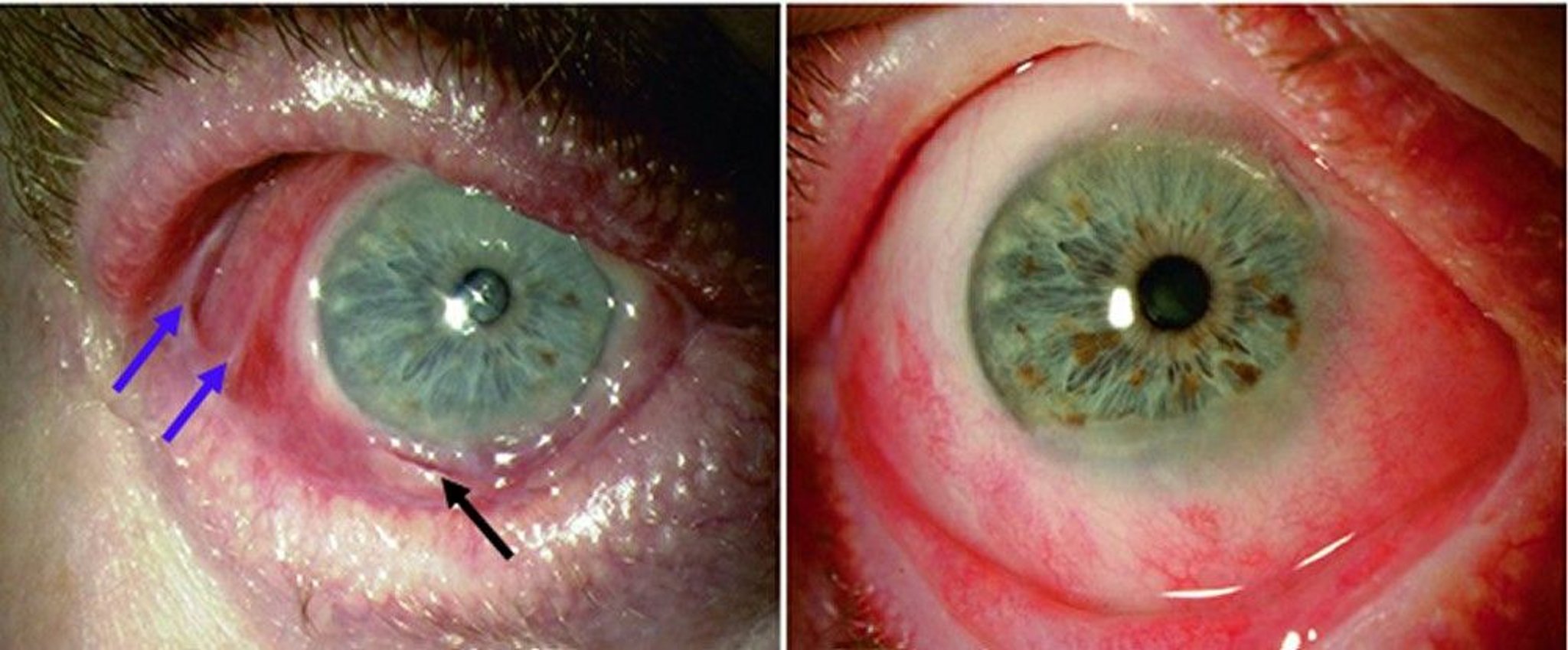
Conjunctival shrinkage with loss of the inferior fornix
Symblephara (adhesions between the palpebral and bulbar conjunctiva)
Trichiasis (in-turning eyelashes)
Corneal epithelial defects and bacterial corneal infection
Corneal neovascularization, opacification, keratinization, and blindness
Oral mucous membrane involvement with ulceration and scarring is common, but skin involvement, characterized by scarring bullae and erythematous plaques, is uncommon.

© Springer Science+Business Media
Diagnosis of Ocular Mucous Membrane Pemphigoid
Ocular examination
Sometimes biopsy
Diagnosis of ocular mucous membrane pemphigoid is suspected clinically in patients with conjunctival scarring as well as corneal changes, symblephara (adhesion of some palpebral conjunctiva to the bulbar conjunctiva), or both (1). Other causes of progressive conjunctival scarring include previous radiation exposure and atopic disease. Therefore, the clinical diagnosis of ocular cicatricial pemphigoid is made when there is progression to a symblepharon without history of local radiation or severe perennial allergic conjunctivitis.
Diagnosis can be confirmed by conjunctival biopsy showing linear antibody deposition on the basement membrane. A negative biopsy result does not rule out the diagnosis.
Diagnosis reference
1. Georgoudis P, Sabatino F, Szentmary N, et al. Ocular Mucous Membrane Pemphigoid: Current State of Pathophysiology, Diagnostics and Treatment. Ophthalmol Ther. 2019;8(1):5-17. doi:10.1007/s40123-019-0164-z
Treatment of Ocular Mucous Membrane Pemphigoid
Topical lubrication
Topical anti-inflammatory therapy (eg, corticosteroids, cyclosporine)Topical anti-inflammatory therapy (eg, corticosteroids, cyclosporine)
Epilation of trichiasis (in-turning eyelashes)
Often systemic therapy with dapsone or immunosuppressantsOften systemic therapy with dapsone or immunosuppressants
In people with ocular mucous membrane pemphigoid, tear substitutes and epilation, cryoepilation, or electroepilation of trichiasis may increase comfort and reduce the risk of ocular infection, secondary corneal scarring, and decreased vision.
For progressive trichiasis, conjunctival scarring, or corneal opacification or for nonhealing corneal epithelial defects, systemic therapy with dapsone (For progressive trichiasis, conjunctival scarring, or corneal opacification or for nonhealing corneal epithelial defects, systemic therapy with dapsone (1) or immunosuppressants (eg, methotrexate, mycophenolate mofetil, cyclophosphamide, intravenous immunoglobulin [IVIG], rituximab) is indicated () or immunosuppressants (eg, methotrexate, mycophenolate mofetil, cyclophosphamide, intravenous immunoglobulin [IVIG], rituximab) is indicated (2).
Patients with nonhealing epithelial defects may also benefit from amniotic membrane transplantation.
Treatment references
1. Branisteanu DC, Stoleriu G, Branisteanu DE, et al. Ocular cicatricial pemphigoid (Review). Exp Ther Med. 2020;20(4):3379-3382. doi:10.3892/etm.2020.8972
2. Georgoudis P, Sabatino F, Szentmary N, et al. Ocular Mucous Membrane Pemphigoid: Current State of Pathophysiology, Diagnostics and Treatment. Ophthalmol Ther. 2019;8(1):5-17. doi:10.1007/s40123-019-0164-z
Key Points
Ocular mucous membrane pemphigoid is a chronic, autoimmune scarring of the conjunctiva with opacification of the cornea.
Findings include symblephara (adhesions between the palpebral and bulbar conjunctiva); trichiasis (in-turning eyelashes); keratoconjunctivitis sicca; corneal neovascularization, opacification, and keratinization; and conjunctival shrinkage and keratinization.
Diagnosis is usually by finding a progressive symblepharon in a patient without a history of local radiation or severe perennial allergic conjunctivitis.
Treatment can include tear substitutes, topical anti-inflammatory therapy, epilation of trichiasis, and often systemic therapy with dapsone or immunosuppressants.Treatment can include tear substitutes, topical anti-inflammatory therapy, epilation of trichiasis, and often systemic therapy with dapsone or immunosuppressants.
Drugs Mentioned In This Article


