

Glaucomas are a group of eye disorders characterized by progressive optic nerve damage in which an important part is a relative increase in intraocular pressure (IOP) that can lead to irreversible loss of vision.
Glaucoma is the second most common cause of blindness worldwide and the second most common cause of blindness in the United States, where it is the leading cause of blindness for Black and Hispanic people. Approximately 4 million Americans (1) and 70 million people worldwide have glaucoma, but only half are aware of it (2). Glaucoma can occur at any age but is 6 times more common among people > age 60.
Glaucomas are categorized as
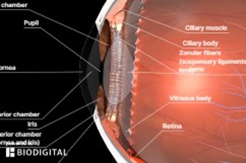
The “angle” refers to the angle formed by the junction of the iris and cornea at the periphery of the anterior chamber (see figure ). The angle is where > 98% of the aqueous humor exits the eye through either the trabecular meshwork and Schlemm canal (the major pathway, particularly in older adults) or the ciliary body face and choroidal vasculature (also called the uveoscleral pathway, which can make up to 50% of outflow in people younger than 30). These outflow pathways are not simply a mechanical filter and drain but instead involve active physiologic processes.
Open-Angle Glaucoma: Classification Based on Mechanisms of Outflow Obstruction*
Type | Means | Examples |
|---|---|---|
Trabecular | ||
Idiopathic | Disorder of extracellular matrix | Corticosteroid-induced glaucoma Juvenile glaucoma Pseudoexfoliation glaucoma |
Obstruction | By red blood cells | Ghost cell glaucoma Hemorrhagic glaucoma |
By macrophages | Hemolytic glaucoma Melanomalytic glaucoma Phacolytic glaucoma | |
By neoplastic cells | Juvenile xanthogranuloma Malignant tumors Nevus of Ota (oculodermal melanocytosis) | |
By pigment particles | Exfoliation syndrome (glaucoma capsulare) Pigmentary glaucoma Uveitis | |
By protein | Lens-induced glaucoma | |
Due to other means | Viscoelastic agents Vitreous hemorrhage | |
Alterations | Due to edema | Alkali burns Iritis or uveitis causing trabeculitis |
Due to trauma | Angle recession | |
Due to intraocular foreign bodies | Chalcosis | |
Posttrabecular | ||
Obstruction of Schlemm canal | By particulate matter or collapse of canal walls | Age-related changes in canal wall Sickled red blood cells Trauma |
Reduced flow in aqueous veins | Due to elevated episcleral venous pressure | Carotid-cavernous fistula Idiopathic episcleral venous pressure elevation Infiltrative ophthalmopathy (thyrotropic exophthalmos) Mediastinal tumors Retrobulbar tumors Superior vena cava obstruction |
* Clinical examples cited; not an inclusive list of glaucomas. | ||
Adapted from Ritch R, Shields MB, Krupin T: The Glaucomas, ed. 2. St. Louis, Mosby, 1996, p. 720; with permission. | ||
Angle-Closure Glaucoma: Classification Based on Mechanisms of Outflow Obstruction*
Mechanism | Examples |
|---|---|
Anterior (pulling mechanism) | |
Contracture of membranes | Iridocorneal endothelial syndrome Neovascular glaucoma Posterior polymorphous dystrophy Surgery (eg, corneal transplant) Trauma (penetrating and nonpenetrating) |
Contracture of inflammatory precipitates | — |
Inflammatory membrane | Fuchs heterochromic iridocyclitis Luetic interstitial keratitis |
Posterior (pushing mechanism) with pupillary block | |
Lens induced | Intumescent lens Subluxation of lens Mobile lens syndrome |
Posterior synechiae | Iris-vitreous block in aphakia Pseudophakia |
Posterior (pushing mechanism) without pupillary block | |
Aqueous misdirection | Ciliary block (malignant glaucoma) |
Cysts of the iris and ciliary body | — |
Forward vitreous shift after lens extraction | — |
Intraocular tumors | |
Lens induced | Intumescent lens Subluxation of lens Mobile lens syndrome |
Large or anterior displaced ciliary body | Plateau iris syndrome |
Uveal edema | After scleral buckling, panretinal photocoagulation, or central retinal vein occlusion |
Retrolenticular tissue contracture | Persistent hyperplastic primary vitreous Retinopathy of prematurity (retrolental fibroplasia) |
* Clinical examples cited; not an inclusive list of glaucomas. | |
Adapted from Ritch R, Shields MB, Krupin T: The Glaucomas, ed. 2. St. Louis, Mosby, 1996, p. 720; with permission. | |
Developmental Abnormalities of the Anterior Chamber Angle Causing Glaucoma: Classification Based on Mechanisms of Outflow Obstruction*
Mechanism | Disorders |
|---|---|
High insertion of peripheral iris | Axenfeld-Rieger syndrome Peters anomaly |
Incomplete development of trabecular meshwork or Schlemm canal | Congenital (infantile) glaucoma Glaucomas associated with other developmental abnormalities (eg, aniridia with congenital onset of glaucoma) |
Fine strands that contract to close angle | Aniridia (juvenile onset of glaucoma) |
* Clinical examples cited; not an inclusive list of the glaucomas. | |
Adapted from Ritch R, Shields MB, Krupin T: The Glaucomas, ed. 2. St. Louis, Mosby, 1996, p. 720; with permission. | |
See tables for classification based on mechanisms of outflow obstruction for open-angle glaucoma, angle-closure glaucoma, and developmental abnormalities of the anterior chamber angle.
Glaucomas are further subdivided into primary (cause of outflow resistance or angle closure is unknown) and secondary (outflow resistance results from a known disorder), accounting for > 20 types of glaucoma in adults.
Aqueous Humor Production and Flow
Most of the aqueous humor, produced by the ciliary body, exits the eye at the angle formed by the junction of the iris and cornea. It exits primarily via the trabecular meshwork and Schlemm canal (pink arrows). A smaller amount drains through the ciliary body face entering the uveoscleral pathway (black arrows). In early adulthood, the ratio of drainage is approximately equal in both pathways. With aging, drainage occurs predominantly through the trabecular meshwork and Schlemm canal. |


References
1. Ehrlich JR, Burke-Conte Z, Wittenborn JS, et al. Prevalence of Glaucoma Among US Adults in 2022. JAMA Ophthalmol. 2024;142(11):1046-1053. doi:10.1001/jamaophthalmol.2024.3884
2. Tham YC, Li X, Wong TY, Quigley HA, Aung T, Cheng CY. Global prevalence of glaucoma and projections of glaucoma burden through 2040: a systematic review and meta-analysis. Ophthalmology. 2014;121(11):2081-2090. doi:10.1016/j.ophtha.2014.05.013
Pathophysiology of Glaucoma
Axons of retinal ganglion cells travel through the optic nerve carrying visual information from the eye to the brain. Damage to these axons causes ganglion cell death with resultant optic nerve atrophy and patchy vision loss. Elevated intraocular pressure (IOP) plays a role in axonal damage, either by direct nerve compression or diminution of blood flow. However, the relationship between externally measured pressure and nerve damage is complicated. Normal IOP ranges from 11 to 21 mm Hg. However, among people whose IOP > 21 mm Hg (ie, ocular hypertension), only approximately 1 to 2% a year, or 10% over 5 years develop glaucoma. Additionally, approximately one-third of patients with glaucoma do not have IOP > 21 mm Hg (known as low-tension glaucoma or normal-tension glaucoma) (1).
One factor that may explain the apparent variability in the correlation between the incidence of glaucoma and measured IOP is that externally measured IOP may not always reflect the true IOP; a thinner than average cornea leads to a higher IOP measurement, while a thicker than average cornea leads to a lower IOP measurement, relative to the true IOP within the eye. It is also likely that there are factors within the optic nerve, such as compromised blood flow, that lead to optic nerve damage.
IOP is determined by the balance of aqueous secretion and drainage. Elevated IOP is caused by inhibited or obstructed outflow, not oversecretion; a combination of factors in the trabecular meshwork (eg, dysregulation of extracellular matrix, cytoskeletal abnormalities) appear to be involved. In open-angle glaucoma, IOP is elevated because outflow is inadequate despite an angle that appears macroscopically unobstructed. In angle-closure glaucoma, IOP is elevated when the peripheral iris mechanically blocks outflow.
Pathophysiology reference
1. Kass MA, Heuer DK, Higginbotham EJ, et al. The Ocular Hypertension Treatment Study: a randomized trial determines that topical ocular hypotensive medication delays or prevents the onset of primary open-angle glaucoma. Arch Ophthalmol. 2002;120(6):701-830. doi:10.1001/archopht.120.6.701
Symptoms and Signs of Glaucoma
Symptoms and signs of glaucoma vary with the type of glaucoma. The defining characteristic of glaucoma is optic nerve damage as evidenced by an abnormal optic disk and certain types of visual field deficits that localize to the portion of the optic nerve that transits through the fenestrated sclera (the lamina cribrosa).
Intraocular pressure (IOP) may be elevated or within the average range. (For techniques of measurement, see Testing.)
Diagnosis of Glaucoma
Characteristic optic nerve changes
Characteristic visual field defects
Exclusion of other causes of visual impairment
Intraocular pressure (IOP) usually > 21 mm Hg (but not required for the diagnosis)
Glaucoma should be suspected in a patient with any of the following:
Abnormal optic nerve on ophthalmoscopy
Elevated IOP
Characteristic visual field defects that localize to the optic nerve
Family history of glaucoma
Such patients (and those with any risk factors) should be referred to an ophthalmologist for a comprehensive examination that includes a thorough history, family history, examination of the optic disks (preferably using a binocular examination technique), formal visual field examination, tonometry (measurement of IOP), measurement of central corneal thickness, optic nerve and/or retinal nerve fiber layer imaging (using optical coherence tomography), and gonioscopy (visualization of the anterior chamber angle with a special mirrored contact lens prism).
Glaucoma is diagnosed when characteristic findings of optic nerve damage are present and other causes of vision disturbance (eg, multiple sclerosis) have been excluded. Elevated IOP makes the diagnosis more likely, but is not essential for making the diagnosis because elevated IOP can occur in the absence of glaucoma. Low-tension (low-pressure) or normal-pressure glaucoma (IOP < 21 mmHg) comprises approximately one-third of all open-angle glaucomas in the United States and is even more common in Asia (1).
Screening


Screening for glaucoma can be done by primary eye clinicians (more commonly an optometrist or ophthalmologist) by frequency-doubling technology (FDT) perimetry to check visual fields and ophthalmoscopic evaluation of the optic nerve. FDT perimetry involves use of a desktop device to rapidly (2 to 3 minutes per eye) screen for visual field abnormalities. Although IOP should be measured, IOP alone has low sensitivity, low specificity, and low positive predictive value as a screening tool for glaucoma. Patients > 40 years and those who have risk factors for open-angle or angle-closure glaucoma should receive a comprehensive eye examination every 1 to 2 years.
Diagnosis reference
1. Leung DYL, Tham CC. Normal-tension glaucoma: Current concepts and approaches-A review. Clin Exp Ophthalmol. 2022;50(2):247-259. doi:10.1111/ceo.14043
Treatment of Glaucoma
Decreasing intraocular pressure (IOP) by using medications or laser or incisional surgery
Patients with characteristic optic nerve and corresponding visual field changes are treated regardless of IOP measurement. Lowering the IOP is the only clinically proven treatment. For chronic adult and juvenile glaucomas, the initial target IOP measurement is at least 20 to 40% below pretreatment readings.
Three methods are available to lower IOP: medications, laser surgery, and incisional surgery. The type of glaucoma determines the appropriate method or methods.
Medications and most laser surgeries (trabeculoplasty) modify the existing aqueous secretion and drainage system.
Traditional incisional surgeries (eg, guarded filtration procedures [trabeculectomy] or glaucoma drainage implant devices [tube shunts]) create a new drainage pathway between the anterior chamber and subconjunctival space. Newer incisional surgeries enhance trabecular or uveoscleral outflow without creating a full-thickness fistula.
Prophylactic IOP lowering in patients with ocular hypertension delays the onset of glaucoma. However, because the rate of conversion from ocular hypertension to glaucoma is low, the decision to treat prophylactically should be individualized, based on the presence of risk factors, magnitude of IOP elevation, and patient factors (ie, preference for medications vs surgery, medication adverse effects). Generally, treatment is recommended for patients with IOP > 30 mm Hg even if the visual field is full and the optic nerve disk appears healthy because the likelihood of damage is significant at that IOP level.
Key Points
Glaucoma is common, often asymptomatic, and is a major cause of blindness worldwide.
Suspect glaucoma if patients have elevated IOP, optic nerve abnormalities on ophthalmoscopy, or a family history of glaucoma.
Do not exclude glaucoma because IOP is not high.
Screen patients > 40 years and patients with risk factors every 1 to 2 years, based mainly on results of ophthalmoscopy and frequency-doubling technology (to assess visual fields).
Treat by decreasing IOP.
Prophylactically decrease IOP if > 30 mm Hg, even if glaucoma is absent.


