


For fertilization (conception), spermatozoa (male haploid gametes) migrate through the cervical canal, the uterine cavity, and into the fallopian tubes. In the ovary, follicles develop and, during ovulation, the dominant follicle releases an oocyte (female haploid gamete). The oocyte enters into the fallopian tube through the fimbriated end, travels through the fallopian tube, and then passes into the uterine cavity. The oocyte becomes a fertilized ovum when a spermatozoon penetrates the outer layers of the oocyte. Fertilization typically occurs while the oocyte is in the fallopian tube.
Female and Male Gametogenesis
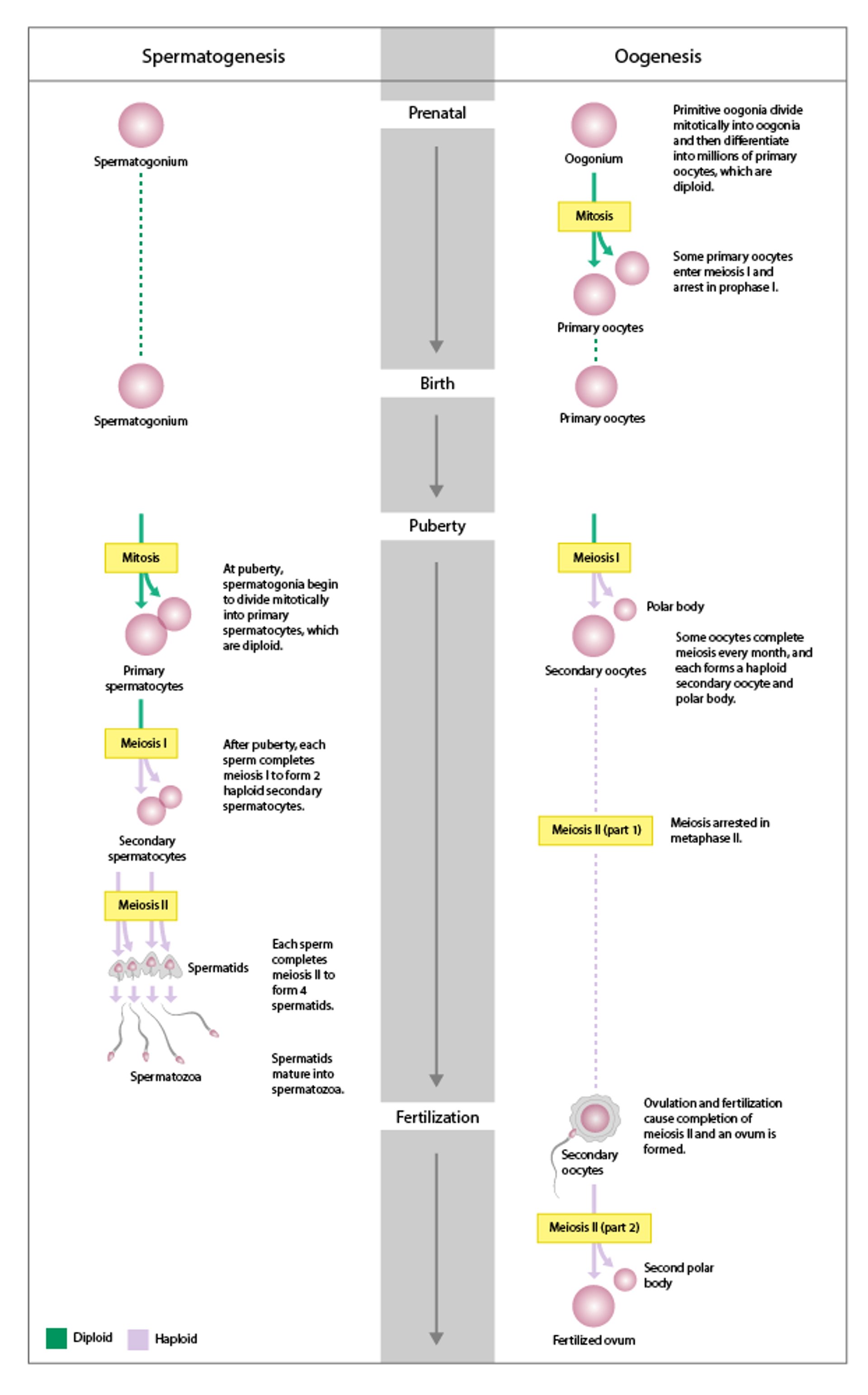
Gametogenesis is the process of development from primordial germ cells to mature gametes: oogenesis in females and spermatogenesis in males. In both females and males, it begins with diploid germ cells that then undergo mitosis, meiosis, and cytodifferentiation into haploid gametes. |

Typically, 1 spermatozoon fertilizes 1 oocyte. However, 2 oocytes may be released and then fertilized by 2 spermatozoa, resulting in a dizygotic (fraternal) twin pregnancy. Higher-order multiple gestation (eg, triplets, quadruplets) may occur if more than 2 oocytes are released and fertilized, but this is rare. Also, more than 1 sperm can penetrate a single oocyte, but this results in an abnormal zygote.
In a woman with 28-day menstrual cycles, ovulation occurs about 14 days after the first day of a menstrual period. At ovulation, cervical mucus becomes less viscid, facilitating rapid movement of spermatozoa to the oocyte. Sperm may remain alive in the reproductive tract for about 3 days after intercourse.
The fertilized ovum (zygote) is diploid. It divides repeatedly as it migrates to the implantation site in the endometrium (usually near the fundus or posterior wall of the uterus). By the time of implantation, the zygote has become a blastocyst, which is a layer of cells around a cavity. The blastocyst wall is 1 cell thick except for the embryonic pole, which is 3 or 4 cells thick. About 6 days after fertilization, the blastocyst implants in the uterine lining; the embryonic pole, which will develop into the embryo, is the first point of implantation.


PIKOVIT / SCIENCE PHOTO LIBRARY
Amniotic sac and placenta
Within 1 or 2 days of implantation, a layer of cells (trophoblast cells) develops around the blastocyst. The progenitor villous trophoblast cell, the stem cell of the placenta, develops along 2 cell lines:
Nonproliferative extravillous trophoblast: These cells penetrate the endometrium, facilitating implantation and anchoring of the placenta.
Syncytiotrophoblast: These cells produce chorionic gonadotropin by day 10 and other trophic hormones shortly thereafter.
An inner layer (amnion) and outer layer (chorion) of membranes develop from the trophoblast; these membranes form the amniotic sac, which contains the conceptus (term used for derivatives of the zygote at any stage—see figure Placenta and Embryo At About 11 4/7 Weeks Gestation). When the sac is formed and the blastocyst cavity closes (by about 10 days), the conceptus is considered an embryo. The amniotic sac fills with fluid and expands with the growing embryo, filling the endometrial cavity by about 12 weeks after conception; then, the amniotic sac is the only cavity remaining in the uterus.
Placenta and Embryo At About 11 4/7 Weeks Gestation
The embryo measures 4.2 cm. |

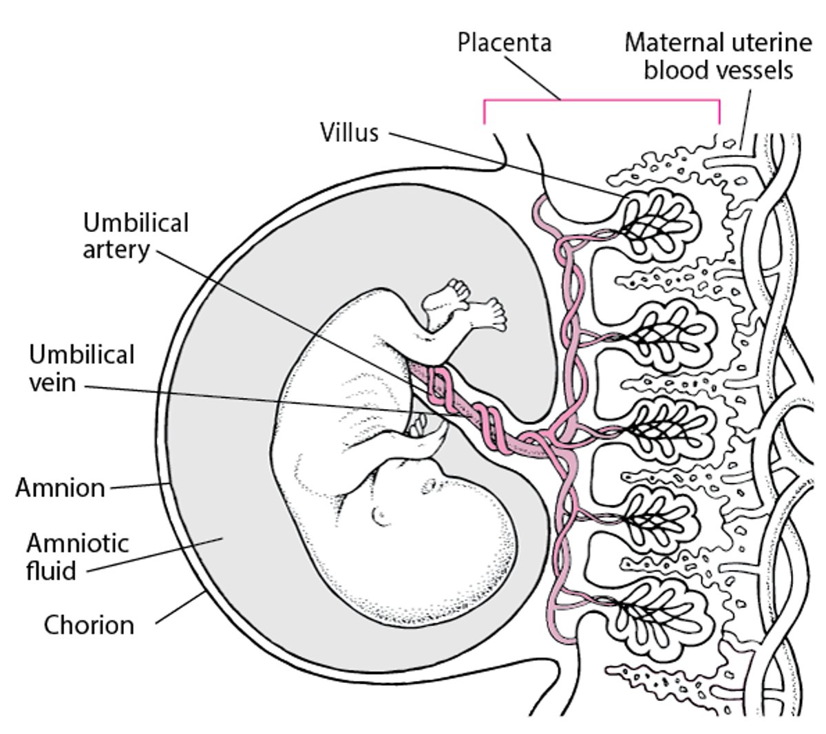
Trophoblast cells develop into cells that form the placenta. The extravillous trophoblast forms villi, which penetrate the uterus. The syncytiotrophoblast covers the villi. The syncytiotrophoblast synthesizes trophic hormones and provides arterial and venous exchange between the circulation of the conceptus and that of the mother.
The placenta is fully formed by week 18 to 20 but continues to grow, weighing about 500 g by term.
Embryo
Around day 10 after fertilization, 3 germ layers (ectoderm, mesoderm, endoderm) are usually distinct in the embryo. Then the primitive streak, which becomes the neural tube, begins to develop.
Around day 16, the cephalad portion of the mesoderm thickens, forming a central channel that develops into the heart and great vessels. The heart begins to pump plasma around day 20, and on the next day, fetal red blood cells (RBCs), which are immature and nucleated, appear. Fetal RBCs are soon replaced by mature RBCs, and blood vessels develop throughout the embryo. Eventually, the umbilical artery and vein develop, connecting the embryonic vessels with the placenta.
Most organs form between 21 and 57 days after fertilization (between 5 and 10 weeks gestation); however, the central nervous system continues to develop throughout pregnancy. Susceptibility to malformations induced by teratogens is highest when organs are forming.


