


(See also Vaginal Bleeding.)
Abnormal uterine bleeding (AUB) is a common issue in reproductive-aged women. Ovulatory dysfunction (anovulation or oligo-ovulation), which occurs most frequently in adolescents and women > 45 years old, is the most common cause of AUB and results in amenorrhea or irregular menses. Other patterns of AUB include heavy menstrual bleeding or intermenstrual bleeding.
Pathophysiology of AUB
In ovulatory dysfunction, during an anovulatory cycle, estrogen is produced, but the corpus luteum does not form in the ovary. Thus, the normal cyclical secretion of progesterone does not occur, and estrogen stimulates the endometrium unopposed. Without opposition by progesterone, the endometrium continues to proliferate, eventually outgrowing its blood supply; it then sloughs incompletely. This causes irregular bleeding, with long intervals without bleeding and/or frequent bleeding that is sometimes profuse and can continue for days or weeks. When this abnormal process occurs repeatedly, endometrial hyperplasia or cancer can develop.
In women with obesity, elevated estrogen levels can disrupt the hormonal balance needed for normal cycles and disrupt ovulation. This can occur thru peripheral conversion of androgens to estrogen leading to negative feedback in the hypothalamic-pituitary-ovarian axis and impair release of gonadotropin-releasing hormone. In addition, obesity changes levels of adipokines (leptin and adiponectin) which can affect hormonal balance. If there is increased androgen production due to insulin resistance or high insulin levels, this can interfere with ovulation. In women with obesity, ovulatory AUB can occur if estrogen levels are high, resulting in amenorrhea alternating with irregular or prolonged bleeding. Increased androgen production due to insulin resistance or high insulin levels can interfere with ovulation.
Irregular menses may also occur in patients with ovulatory cycles if the cycle is prolonged. This may occur if there is a short follicular phase and luteal phase dysfunction (due to inadequate progesterone stimulation of the endometrium); a rapid decrease in estrogen before ovulation can cause spotting. This may also occur if progesterone secretion is prolonged during the luteal phase of the menstrual cycle. Irregular shedding of the endometrium results, probably because estrogen levels remain low, near the threshold for bleeding (as occurs during menses).
Other mechanisms of irregular or intermenstrual uterine bleeding are nonhormonal causes of endometrial bleeding (endometritis, endometrial polyps, endometrial hyperplasia or cancer, submucosal leiomyomas).
Heavy menstrual bleeding (cyclic bleeding with increased volume) may be caused by structural lesions that interfere with myometrial function (leiomyomas, adenomyosis) or coagulopathy.
Complications
Chronic heavy or prolonged uterine bleeding may cause iron deficiency anemia. Acute severe hemorrhage may also occur.
If AUB is due to ovulatory dysfunction, infertility may also be present.
Etiology of AUB
Causes of AUB in nonpregnant women of reproductive age may be classified as structural or nonstructural to aid in identification of the cause and guide treatment. The PALM-COEIN classification system may be used (1). PALM-COEIN is a mnemonic for the structural causes (PALM) and nonstructural (COEIN) causes of abnormal bleeding (see figure PALM-COEIN Classification System).
PALM-COEIN Classification System

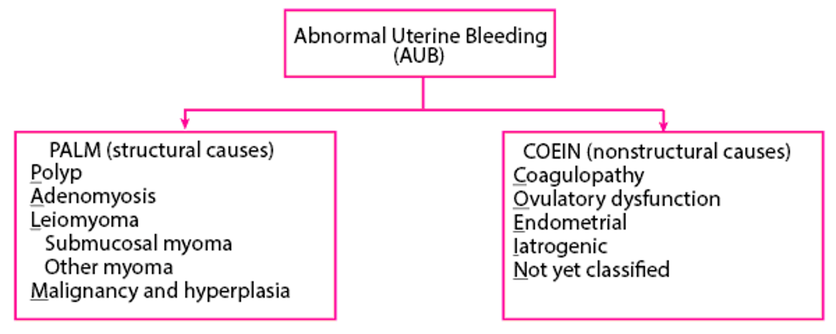
AUB due to ovulatory dysfunction (AUB-O) is the most common type of nonstructural AUB and the most common cause overall. AUB-O can result from any disorder or condition that causes anovulation or oligo-ovulation (irregular or infrequent ovulation—see table Some Etiologies of Ovulatory Dysfunction). Causes of ovulatory dysfunction include:
Perimenarche or perimenopause
Androgen excess (particularly due to polycystic ovary syndrome)
Pituitary dysfunction (eg, hyperprolactinemia cause by pituitary adenoma, pituitary irradiation),
Idiopathic (sometimes occurring when gonadotropin levels are normal)
Hypothalamic dysfunction may result in decreased GnRH production, which in turn may cause decreased gonadotropin production. A common cause is insufficient energy intake due to dietary restriction, undernutrition, or strenuous exercise or emotional stress. Women with amenorrhea due to hypothalamic dysfunction have lower levels of serum leptin (an anorectic hormone produced by fat cells); lower levels may contribute to decreased gonadotropin production (2).
During perimenopause, AUB-O may be an early sign of ovarian insufficiency; follicles are still developing but, despite increasing levels of follicle-stimulating hormone (FSH), do not produce enough estrogen to trigger ovulation.
Some data suggests that endometriosis is associated with AUB-O, but the mechanisms are unknown and study results are inconsistent (2).
Other causes of ovulatory dysfunction are systemic disease (eg, liver or kidney disease, Cushing syndrome, autoimmune diseases [eg, lupus]). Systemic illnesses can disrupt the hypothalamic-pituitary-ovarian axis that regulates the menstrual cycle, thus impacting hormone production and disrupting ovulation.
Significant physical or emotional stress or poor nutrition are typical causes of hypothalamic amenorrhea, but some patients with these factors have oligo-ovulation and, thus, oligomenorrhea.
Other nonstructural causes of AUB include:
Coagulopathy
Endometrial factors (eg, endometritis)
Iatrogenic (eg, breakthrough bleeding on hormonal contraceptives)
Structural types of AUB include:
Endometrial polyps
Adenomyosis
Leiomyoma (uterine fibroids)
Endometrial hyperplasia or uterine cancer
Etiology references
1. Munro MG, Critchley HOD, Fraser IS; FIGO Menstrual Disorders Committee. The two FIGO systems for normal and abnormal uterine bleeding symptoms and classification of causes of abnormal uterine bleeding in the reproductive years: 2018 revisions [published correction appears in Int J Gynaecol Obstet. 2019 Feb;144(2):237. doi: 10.1002/ijgo.12709.]. Int J Gynaecol Obstet. 2018;143(3):393-408. doi:10.1002/ijgo.12666
2. Santulli P, Tran C, Gayet V, et al. Oligo-anovulation is not a rarer feature in women with documented endometriosis. Fertil Steril. 2018;110(5):941-948. doi:10.1016/j.fertnstert.2018.06.012
Symptoms and Signs of AUB
Compared with typical menses, AUB may be characterized by (see table Normal Menstrual Parameters [1]):
Increased frequency (menses < 24 days apart)
Irregular (cycle frequency varies ≥ 8 to 10 days)
Prolonged bleeding (> 8 days)
Increased volume of blood loss (> 80 mL [or by patient report of volume]) during menses (heavy menstrual bleeding)
Intermenstrual bleeding
Normal Menstrual Parameters*
Parameters | Normal Values | Notes |
|---|---|---|
Frequency | ≥ 24 to ≤ 38 days | Cycle length is defined as the number of days from first day of one menstrual period to first day of the next. |
Regularity | ≤ 7 to 9 days | Regularity is defined as variation of cycle frequency between the shortest and longest cycles. |
Duration | ≤ 8 days of bleeding per cycle | — |
Volume of bleeding | < 80 mL Clinically, patient's description of bleeding as normal volume | Clinically, precise measurement bleeding volume is not feasible. Assessment of bleeding volume is based on the patient's description (light, normal, heavy). Clinicians sometimes estimate by asking how many pads or tampons are saturated over time (heavy bleeding is likely if patients saturate a pad or tampon within 3 hours or less and/or if they pass blood clots larger than 1 inch [2.5 cm] in diameter). |
* Based on Munro MG, Critchley HOD, Fraser IS; FIGO Menstrual Disorders Committee. The two FIGO systems for normal and abnormal uterine bleeding symptoms and classification of causes of abnormal uterine bleeding in the reproductive years: 2018 revisions [published correction appears in Int J Gynaecol Obstet. 2019 Feb;144(2):237. doi: 10.1002/ijgo.12709.]. Int J Gynaecol Obstet. 2018;143(3):393-408. doi:10.1002/ijgo.12666 | ||
Patients with ovulatory cycles usually have menstrual cycles that are occur at a normal frequency and are regular, but they may have heavy menstrual bleeding or intermenstrual bleeding. In addition to a regular menses, symptoms that suggest a patient has ovulatory cycles include molimina (eg, cyclic breast tenderness, premenstrual bloating, or mood changes) and midcycle cramping pain (mittelschmerz). Daily body temperature (basal body temperature) increases slightly after ovulation and then decreases after the next menstrual cycle begins.
Patients with AUB-O (ovulatory dysfunction) have amenorrhea or uterine bleeding that occurs at unpredictable times, may vary widely in volume, and is not accompanied cyclic changes in basal body temperature.
Symptoms and signs reference
1. Munro MG, Critchley HOD, Fraser IS; FIGO Menstrual Disorders Committee. The two FIGO systems for normal and abnormal uterine bleeding symptoms and classification of causes of abnormal uterine bleeding in the reproductive years: 2018 revisions [published correction appears in Int J Gynaecol Obstet. 2019 Feb;144(2):237. doi: 10.1002/ijgo.12709.]. Int J Gynaecol Obstet. 2018;143(3):393-408. doi:10.1002/ijgo.12666
Diagnosis of AUB
Menstrual history
Pregnancy test
Hormone blood tests
Blood tests for anemia
Sometimes, blood tests for a bleeding disorder
Pelvic imaging studies, usually transvaginal ultrasound
Sometimes procedures (endometrial sampling or hysteroscopy)
Pregnancy should be excluded, even in young adolescents and perimenopausal women.
The pattern of abnormal uterine bleeding often suggests possible causes (eg, regular cycles with prolonged or excessive bleeding suggest structural abnormalities; irregular bleeding or amenorrhea is often due to ovulatory dysfunction) and helps guide the choice of laboratory or imaging tests.
Coagulation disorders should be considered in adolescents who have anemia or require hospitalization for bleeding and in patients with a family history or other risk factors for coagulation disorders or with a history or unexplained easy or severe bleeding or bruising (1, 2).
Laboratory testing
Pregnancy testing is sometimes done with urine human chorionic gonadotropin (hCG) initially. If this is negative, a serum quantitative hCG should be done. If positive, the patient should be evaluated for bleeding during pregnancy.
Anemia may be severe in women who regularly have heavy and/or prolonged periods. CBC and ferritin are measured to evaluate for iron deficiency anemia .
For adolescents with heavy uterine bleeding or patients with risk factors for or a personal or family medical history suggestive of a bleeding disorder, the platelet count from the CBC may be informative, and other initial blood tests include: prothrombin time and partial thromboplastin time (fibrinogen or thrombin time are optional and bleeding time is not necessary). Based on the initial test results, additional tests may be sent for von Willebrand disease or other bleeding disorders.
To determine whether a patient is anovulatory or ovulatory, some clinicians measure serum progesterone levels during the luteal phase (after day 14 of a normal menstrual cycle or after basal body temperature increases, as occurs during this phase). A level of ≥ 3 ng/mL (≥ 9.75 nmol/L) suggests that ovulation has occurred. Another option is for patients to use a home test kit for urine LH levels, which are measured daily for several days beginning at or after cycle day 9.
Testing for endocrinologic etiologies includes TSH and prolactin levels (even when galactorrhea is absent), because thyroid disorders and hyperprolactinemia are common causes of AUB.
Other laboratory tests may be done depending on results of the history and physical examination and include the following:
Testosterone and dehydroepiandrosterone sulfate (DHEAS) levels if polycystic ovary syndrome is suspected
Serum glucose and lipid levels, blood pressure, and body mass index if polycystic ovary syndrome is suspected
Follicle-stimulating hormone (FSH) and estradiol levels if primary ovarian insufficiency is possible
Tests done to exclude other causes of vaginal bleeding include:
A cervical cancer screening test (Papanicolaou [Pap] test and/or human papillomavirus [HPV] test) if the patient is due for routine screening or a biopsy if a suspicious cervical lesion is seen during a pelvic examination
Testing for Neisseria gonorrhea and Chlamydia species if pelvic inflammatory disease or cervicitis is suspected
Imaging studies
Transvaginal ultrasound is part of the evaluation in most patients with AUB. Specifically, it is done if women have any of the following:
Suspected structural lesion based on bleeding pattern, other symptoms, or pelvic examination findings
Inadequate pelvic examination
Premenopausal women ≥ 45 years old
Postmenopausal women
Risk factors for endometrial cancer (eg, obesity, diabetes, hypertension, polycystic ovary syndrome, chronic eugonadal anovulation, other conditions associated with prolonged unopposed estrogen exposure)
Bleeding that continues despite use of empiric hormone therapy
Transvaginal ultrasound can detect structural abnormalities, including endometrial thickening, endometrial polyps, fibroids, other uterine masses, adenomyosis, and ovarian or fallopian tube abnormalities.
If focal thickening is detected on ultrasound, further testing may be needed to identify smaller intrauterine masses (eg, small endometrial polyps, submucous myomas).
For premenopausal women, ultrasound assessment of endometrial thickness is not used for evaluation for endometrial neoplasia, because the endometrial thickness varies across the menstrual cycle (3):
During menstruation: 2 to 4 mm
Early proliferative phase (cycle days 6 to 14): 5 to 7 mm
Late proliferative phase: ≤ 11 mm
Secretory phase: 7 to 16 mm
In addition, measurement of endometrial thickness to detect endometrial cancer is not always accurate in Black women, thus endometrial sampling may be needed for early cancer detection (4).
For some patients with a first episode of postmenopausal bleeding, measurement of endometrial thickness (endometrial stripe) with transvaginal ultrasound may be used as a first-line test to evaluate for endometrial neoplasia (hyperplasia or cancer) (5). However, ultrasound evaluation is not sufficient, and endometrial sampling is required in patients with:
Risk factors for endometrial cancer (eg, obesity, current or recent tamoxifen therapy)Risk factors for endometrial cancer (eg, obesity, current or recent tamoxifen therapy)
Persistent or recurrent bleeding
Endometrial thickness > 4 to 5 mm determined during ultrasound (as a follow-up test)
Sonohysterography (ultrasound after saline is infused into the uterus) is useful in evaluating such abnormalities; it can be used to determine whether hysteroscopy, a more invasive test, is indicated and to plan resection of intrauterine masses. Or hysteroscopy may be done without sonohysterography.
MRI provides detailed images that are useful in planning surgery but is expensive and is not the first-line imaging test for patients with AUB.
Endometrial sampling
Endometrial sampling is usually recommended to exclude hyperplasia or cancer in women with any of the following:
Premenopausal women aged ≥ 45 years
Age < 45 years with one or more risk factors for endometrial cancer
Abnormal bleeding that is persistent or recurs after a normal initial evaluation and despite treatment
Postmenopausal patients with risk factors for uterine cancer or transvaginal ultrasound with abnormal findings (endometrial thickness > 4 to 5 mm or with focal or irregular endometrial thickening)
Inconclusive ultrasound findings in a patient with suspected endometrial neoplasia
Endometrial sampling may be done as an office endometrial biopsy or a dilation and curettage procedure. In a meta-analysis of studies including 1607 women, the sensitivity of endometrial sampling methods for detecting endometrial cancer was 78% and for atypical endometrial hyperplasia was 76% (6). Most endometrial biopsy specimens contain proliferative or dyssynchronous endometrium, which confirms anovulation because no secretory endometrium is found.
Directed biopsy (with hysteroscopy) may be done to visualize the endometrial cavity directly and do targeted biopsies of focal endometrial abnormalities.
Contraindications to endometrial sampling procedures include pregnancy, uncontrolled bleeding disorders, and acute pelvic infection.
Diagnosis references
1. Screening and Management of Bleeding Disorders in Adolescents With Heavy Menstrual Bleeding: ACOG COMMITTEE OPINION, Number 785. Obstet Gynecol. 2019;134(3):e71-e83. doi:10.1097/AOG.0000000000003411
2. Committee on Practice Bulletins—Gynecology. Practice bulletin no. 128: diagnosis of abnormal uterine bleeding in reproductive-aged women. Obstet Gynecol. 2012;120(1):197-206. doi:10.1097/AOG.0b013e318262e320
3. Committee on Practice Bulletins—Gynecology. Practice bulletin no. 128: diagnosis of abnormal uterine bleeding in reproductive-aged women. Obstet Gynecol. 2012 (reaffirmed 2024);120(1):197-206. doi:10.1097/AOG.0b013e318262e320
4. Doll KM, Pike M, Alson J, et al. Endometrial Thickness as Diagnostic Triage for Endometrial Cancer Among Black Individuals [published correction appears in JAMA Oncol. 2025 Jan 23. doi: 10.1001/jamaoncol.2024.6781.]. JAMA Oncol. 2024;10(8):1068-1076. doi:10.1001/jamaoncol.2024.1891
5. ACOG Committee Opinion No. 734: The Role of Transvaginal Ultrasonography in Evaluating the Endometrium of Women With Postmenopausal Bleeding. Obstet Gynecol. 2018 (reaffirmed 2023);131(5):e124-e129. doi:10.1097/AOG.0000000000002631
6. Sakna NA, Elgendi M, Salama MH, Zeinhom A, Labib S, Nabhan AF. Diagnostic accuracy of endometrial sampling tests for detecting endometrial cancer: a systematic review and meta-analysis. BMJ Open. 2023;13(6):e072124. Published 2023 Jun 23. doi:10.1136/bmjopen-2023-072124
Treatment of AUB
Medications to control bleeding, usually a nonsteroidal anti-inflammatory drug (NSAID), tranexamic acid, or hormone therapyMedications to control bleeding, usually a nonsteroidal anti-inflammatory drug (NSAID), tranexamic acid, or hormone therapy
Iron (oral or intravenous) for iron deficiency anemia, if present
Sometimes progestin therapy for endometrial hyperplasia
Sometimes a procedure to treat structural lesions (eg, hysteroscopic myomectomy, uterine fibroid embolization)
Hysterectomy for persistent bleeding (depending on patient preference and other treatment options) or for endometrial hyperplasia or cancer
Medications
Nonhormonal medications for abnormal uterine bleeding have fewer risks and adverse effects than hormone therapy and can be given intermittently, when bleeding occurs. They are used mainly to treat women who desire pregnancy, wish to avoid hormone therapy, or have heavy regular bleeding (menorrhagia). Options include:
Nonsteroidal anti-inflammatory drugs (NSAIDs), which reduce bleeding by approximately 25 to 35% and relieve dysmenorrhea by reducing prostaglandin levels (1)
Tranexamic acid is an antifibrinolytic agent that inhibits Tranexamic acid is an antifibrinolytic agent that inhibitsplasminogen activation that, when given for up to 5 days during menstruation, reduces menstrual blood loss by approximately 50% (2, 3)
Hormone therapy (eg, estrogen/progestin contraceptives, progestins, a long-acting progestin-releasing intrauterine device [IUD]) is often tried first in women who want contraception or who are perimenopausal. This therapy does the following:
Suppresses endometrial development
Reestablishes predictable bleeding patterns
Decreases menstrual flow
Contraceptive hormone therapy is continued for as long as the patient wishes to use contraception. Once bleeding has been controlled for a few months, patients may choose to continue hormone therapy or to stop therapy to see if AUB is still present.
Combined estrogen/progestin oral contraceptives (COCs) are commonly given. COCs, used cyclically or continuously, can control abnormal uterine bleeding due to ovulatory dysfunction. Also, for women with heavy menstrual bleeding (eg, due to fibroids or adenomyosis), COCs decrease menstrual volume. Progestin-only oral contraceptives do not control heavy bleeding. Benefits of COCs include
Decreasing menstrual blood loss by approximately 35 to 70% (4)
Decreasing dysmenorrhea
Decreasing risk of uterine and ovarian cancer
Risks of an OC depend on the type of OC, dose, duration of use, and patient factors.
A progestogen may be used in the following cases:
Estrogen is contraindicated (eg, for patients with cardiovascular risk factors or prior deep vein thrombosis).
Estrogen is declined by the patient.
Withdrawal bleeding may be more predictable with cyclic progestin therapy (medroxyprogesterone acetate 10 mg/day orally or norethindrone acetate 2.5 to 5 mg/day orally) given for 21 days a month than with a COC. Cyclic natural (micronized) progesterone 200 mg/day for 21 days a month may be used, particularly if pregnancy is possible; however, it may cause drowsiness and does not decrease blood loss as much as a progestin.Withdrawal bleeding may be more predictable with cyclic progestin therapy (medroxyprogesterone acetate 10 mg/day orally or norethindrone acetate 2.5 to 5 mg/day orally) given for 21 days a month than with a COC. Cyclic natural (micronized) progesterone 200 mg/day for 21 days a month may be used, particularly if pregnancy is possible; however, it may cause drowsiness and does not decrease blood loss as much as a progestin.
If patients using noncontraceptive cyclic progestins or progesterone wish to prevent pregnancy, contraception should be used. Contraceptive progestin options include If patients using noncontraceptive cyclic progestins or progesterone wish to prevent pregnancy, contraception should be used. Contraceptive progestin options include
Levonorgestrel-releasing IUD:Levonorgestrel-releasing IUD: It reduces menstrual volume by approximately 70 to 95% (4), provides contraception, and relieves dysmenorrhea; the expulsion rate is higher if fibroids are present (5).
Depot medroxyprogesterone acetate:Depot medroxyprogesterone acetate: They cause amenorrhea and provide contraception but may cause irregular spotting and reversible bone loss.
Other medications are used for particular indications or in patient who cannot use more common treatments.
Gonadotropin-releasing hormone (GnRH) agonists or antagonists suppress ovarian hormone production and cause amenorrhea. They are used to shrink fibroids or stop heavy bleeding prior to surgical treatment. However, their hypoestrogenic adverse effects (eg, osteoporosis, menopausal symptoms) limit their use to 6 months. If long-term use is required, add-back low-dose estrogen and progestin therapy may be given.
GnRH agonists initially cause a surge in luteinizing hormone and follicle stimulating hormone, resulting in an increase in estradiol (6, 7). Amenorrhea is induced after 7 to 14 days. These medications are available as intramuscular injections, nasal sprays, or implants.
GnRH antagonists do not induce an initial estradiol flare and rapidly and reversibly suppress gonadotropins and ovarian sex hormones, which reduces heavy bleeding within a few days. Oral GnRH antagonists are available, some of which are combined with estrogen and progestin as add-back therapy.
Selective progesterone receptor modulatorsSelective progesterone receptor modulators impede cellular proliferation of leiomyoma cells. Use of ulipristal acetate is limited due to potential adverse liver effects. Mifepristone has also been used for symptom relief. impede cellular proliferation of leiomyoma cells. Use of ulipristal acetate is limited due to potential adverse liver effects. Mifepristone has also been used for symptom relief.
DanazolDanazol reduces menstrual blood loss (by causing endometrial atrophy) but is not frequently used because it has many androgenic adverse effects, which may be lessened by using lower doses or a vaginal formulation. To be effective, danazol must be taken continuously, usually for about 3 months. It is usually used only when other forms of therapy are contraindicated. reduces menstrual blood loss (by causing endometrial atrophy) but is not frequently used because it has many androgenic adverse effects, which may be lessened by using lower doses or a vaginal formulation. To be effective, danazol must be taken continuously, usually for about 3 months. It is usually used only when other forms of therapy are contraindicated.
If pregnancy is desired and bleeding is not heavy, ovulation induction with clomipheneclomiphene may be used.
Procedures
Hysteroscopy with dilation and curettage (D&C) may be therapeutic as well as diagnostic; it may be the treatment of choice when uterine bleeding volume is severe or when hormone therapy is ineffective. Structural causes such as polyps or fibroids may be identified or removed during hysteroscopy. This procedure typically decreases bleeding for a few months; a rare complication is endometrial scarring (Asherman syndrome).
Endometrial ablation (eg, rollerball, resectoscopic, radiofrequency, thermal, or cryotherapy) decreases volume of bleeding in the majority of patients. Ablation is minimally invasive. Ablation may be repeated if heavy bleeding recurs after ablation is initially effective. If this treatment does not control bleeding or if bleeding continues to recur, the cause may be adenomyosis and thus is not abnormal uterine bleeding due to ovulatory dysfunction. Endometrial ablation does not prevent pregnancy. Pregnancy rates may be as high as 5% after ablation. Ablation causes scarring, which may make sampling the endometrium difficult later.
Treatment of fibroids includes (5):
Minimally invasive procedures, the most commonly used is uterine artery embolization, a procedure in which fibroids are visualized fluoroscopically and emboli are injected through femoral artery catheters to occlude blood supply to the fibroids. Other options include magnetic resonance guided focused ultrasound, radiofrequency ablation, and other techniques in testing such as high intensity focused ultrasound energy.
Myomectomy (removal of uterine fibroids) may be done hysteroscopically for submucosal fibroids or laparoscopically or via laparotomy for intramural or subserosal fibroids.
The location, size, number of fibroids, and coexistent adenomyosis should be taken into account as well as desire for fertility or preservation of uterus.
Hysterectomy, laparoscopic, abdominal, or vaginal, may be recommended for patients who decline hormone therapy or who, despite other treatments, have symptomatic anemia or poor quality of life caused by persistent, irregular bleeding.
Management of acute uterine bleeding
Uterine bleeding can sometimes be acute with rapid loss of a large volume of blood. Emergency measures are similar to management of hemorrhage from other sites, including: close monitoring of vital signs, stabilizing the patient hemodynamically with IV crystalloid fluid, and blood products, as needed. Pregnancy must be excluded. In addition to CBC, labs should be drawn to evaluate for disseminated intravascular coagulation.
Procedures for urgent management include: intrauterine tamponade (eg, inserting a bladder catheter into the uterus and inflating it with 30 to 60 mL of fluid), D&C, or uterine artery embolization. Once patients are stable, hormone therapy is used to control bleeding. Medications may also be used alone or in combination with procedures, including (8):
High-dose estrogen-progestin oral contraceptives (OC): monophasic OCs that contain 35 mcg ethinyl estradiol and progestin, 3 times daily for 7 days High-dose estrogen-progestin oral contraceptives (OC): monophasic OCs that contain 35 mcg ethinyl estradiol and progestin, 3 times daily for 7 days
High-dose oral progestins: medroxyprogesterone acetate 20 mg, 3 times daily for 7 days High-dose oral progestins: medroxyprogesterone acetate 20 mg, 3 times daily for 7 days
Antifibrinolytics: tranexamic acid 1.3 g orally or 10 mg/kg IV (maximum 600 mg), 3 times daily for 5 daysAntifibrinolytics: tranexamic acid 1.3 g orally or 10 mg/kg IV (maximum 600 mg), 3 times daily for 5 days
Intravenous estrogen: conjugated estrogens 25 mg IV, every 4 to 6 hours for a total of 4 doses Intravenous estrogen: conjugated estrogens 25 mg IV, every 4 to 6 hours for a total of 4 doses
High-dose steroid estrogen, progestin, or tranexamic acid therapy is associated with an increased risk of thrombosis, so patient selection, education, and monitoring are important.High-dose steroid estrogen, progestin, or tranexamic acid therapy is associated with an increased risk of thrombosis, so patient selection, education, and monitoring are important.
After an acute management regimen is completed, patients are typically given a daily estrogen-progestin oral contraceptive, which may be continued until bleeding has been controlled for a few months.
If bleeding cannot be controlled with conservative measures and the patient is unstable, hysterectomy is an option. However, all attempts are made to avoid this, particularly in patient with plans for future childbearing.
Treatment references
1. Bofill Rodriguez M, Lethaby A, Farquhar C. Non-steroidal anti-inflammatory drugs for heavy menstrual bleeding. Cochrane Database Syst Rev. 2019;9(9):CD000400. Published 2019 Sep 19. doi:10.1002/14651858.CD000400.pub4
2. Bonnar J, Sheppard BL. Treatment of menorrhagia during menstruation: randomised controlled trial of ethamsylate, mefenamic acid, and tranexamic acid. . Treatment of menorrhagia during menstruation: randomised controlled trial of ethamsylate, mefenamic acid, and tranexamic acid.BMJ. 1996;313(7057):579-582. doi:10.1136/bmj.313.7057.579
3. Lukes AS, Moore KA, Muse KN, et al. Tranexamic acid treatment for heavy menstrual bleeding: a randomized controlled trial. . Tranexamic acid treatment for heavy menstrual bleeding: a randomized controlled trial.Obstet Gynecol. 2010 (reaffirmed 2024);116(4):865-875. doi:10.1097/AOG.0b013e3181f20177
4. Matteson KA, Rahn DD, Wheeler TL 2nd, et al. Nonsurgical management of heavy menstrual bleeding: a systematic review. Obstet Gynecol. 2013;121(3):632-643. doi:10.1097/AOG.0b013e3182839e0e
5. Vannuccini S, Petraglia F, Carmona F, Calaf J, Chapron C. The modern management of uterine fibroids-related abnormal uterine bleeding. Fertil Steril. 2024;122(1):20-30. doi:10.1016/j.fertnstert.2024.04.041
6. Schlaff WD, Ackerman RT, Al-Hendy A, et al: Elagolix for heavy menstrual bleeding in women with uterine fibroids. : Elagolix for heavy menstrual bleeding in women with uterine fibroids.N Engl J Med 382 (4):328–340, 2020. doi: 10.1056/NEJMoa1904351
7. de Lange ME, Huirne JAF: Linzagolix: An oral gonadotropin-releasing hormone receptor antagonist treatment for uterine fibroid-associated heavy menstrual bleeding. Lancet 400 (10356):866–867, 2002. doi: 10.1016/S0140-6736(22)01781-0
8. ACOG committee opinion no. 557: Management of acute abnormal uterine bleeding in nonpregnant reproductive-aged women. Obstet Gynecol. 2013;121(4):891-896. doi:10.1097/01.AOG.0000428646.67925.9a
Guidelines for Abnormal Uterine Bleeding
The following is a list of professional medical society or government clinical practice guidelines regarding this medical issue (this is not a comprehensive list):
American College of Obstetricians and Gynecologists (ACOG) Practice Bulletin. Diagnosis of Abnormal Uterine Bleeding in Reproductive-Aged Women. 2012 (reaffirmed 2024).
ACOG Committee Opinion. Management of Acute Abnormal Bleeding in Nonpregnant Reproductive-Aged Women. 2013 (reaffirmed 2024).
ACOG Practice Bulletin. Management of Abnormal Uterine Bleeding Associated With Ovulatory Dysfunction. 2013 (reaffirmed 2022).
ACOG Practice Bulletin. Management of Symptomatic Uterine Leiomyomas. 2021.
ACOG Committee Opinion. Screening and Management of Bleeding Disorders in Adolescents With Heavy Menstrual Bleeding. 2019; ACOG Committee Opinion. Screening and Management of Bleeding Disorders in Adolescents With Heavy Menstrual Bleeding: Correction (2023)
ACOG Committee Opinion. The Role of Transvaginal Ultrasonography in Evaluating the Endometrium of Women With Postmenopausal Bleeding. 2018 (reaffirmed 2023).
Key Points
Abnormal uterine bleeding is a common medical issue; ovulatory dysfunction is the most common cause of abnormal uterine bleeding.
Test for treatable causes of bleeding and iron deficiency anemia; tests may include a pregnancy test, CBC and ferritin, measurement of hormone levels (TSH, prolactin, progesterone), and often ultrasound, office hysteroscopy, and endometrial sampling.Test for treatable causes of bleeding and iron deficiency anemia; tests may include a pregnancy test, CBC and ferritin, measurement of hormone levels (TSH, prolactin, progesterone), and often ultrasound, office hysteroscopy, and endometrial sampling.
In women at risk, do endometrial sampling to check for endometrial hyperplasia or cancer.
If medications are needed to control bleeding, treat with NSAIDs, tranexamic acid, estrogen-progestin OCs, levonorgestrel-releasing IUDs, gonadotrophin agonists or antagonists, or other hormones.If medications are needed to control bleeding, treat with NSAIDs, tranexamic acid, estrogen-progestin OCs, levonorgestrel-releasing IUDs, gonadotrophin agonists or antagonists, or other hormones.
Treat structural lesions or bleeding that does not respond to medications with a procedure (eg, hysteroscopy, endometrial ablation, hysterectomy).
Drugs Mentioned In This Article


