


Topic Resources

(See also Overview of Congenital Gastrointestinal Anomalies.)
The estimated incidence of gastroschisis is 1 in 2500 live births (1). It is more common than omphalocele.
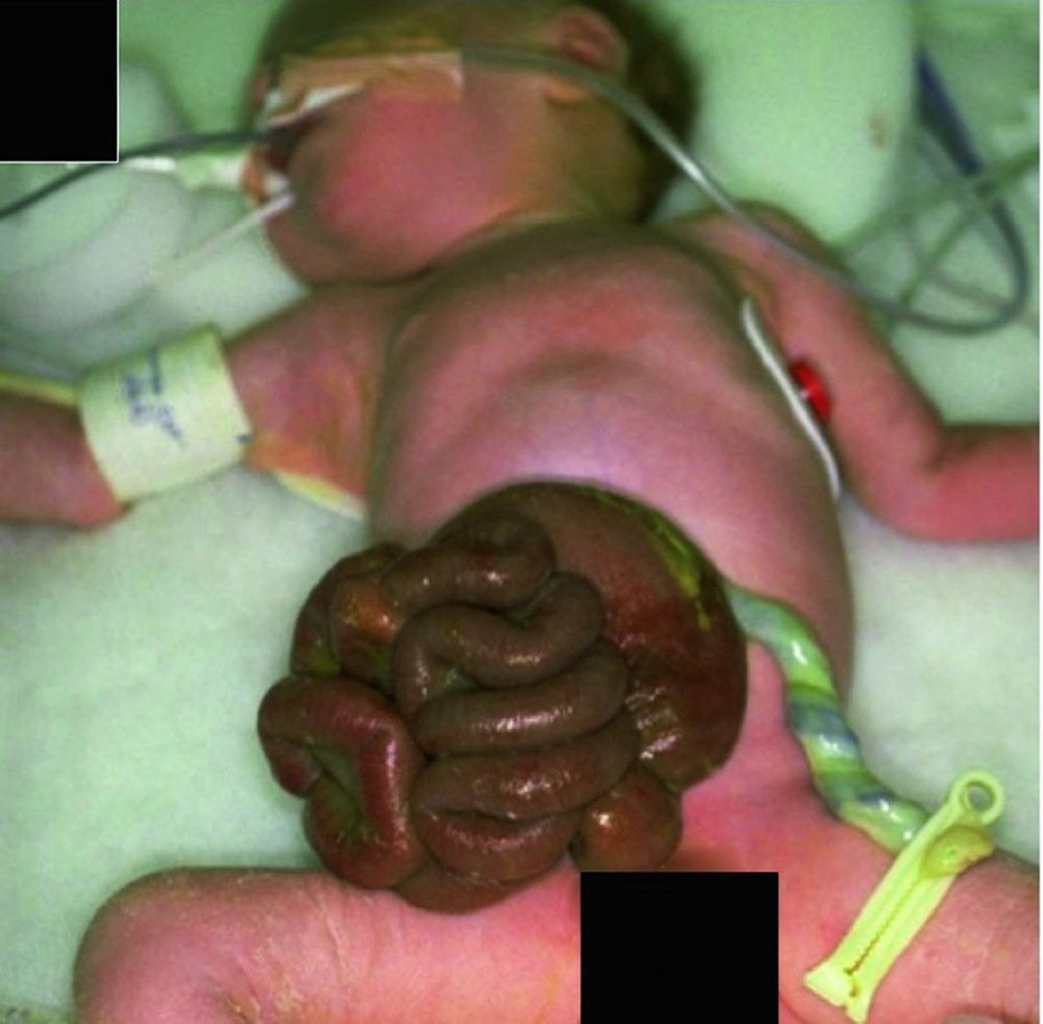
In gastroschisis, the defect is usually to the right of the umbilical cord insertion. Location of the defect differs from omphalocele. In omphalocele, the midline defect is at the base of the umbilicus.
In gastroschisis, unlike omphalocele, there is no membranous covering over the intestine, which is markedly edematous and erythematous and is often enclosed in a fibrin mat. These findings indicate long-standing inflammation due to the intestine being directly exposed to amniotic fluid (ie, chemical peritonitis), which can ultimately result in fibrosis, dysmotility, or obstruction.
Infants with gastroschisis have low incidence of associated congenital anomalies (10 to 15%) other than gastrointestinal abnormalities such as malrotation and intestinal atresia, which occur in approximately 25% of infants (2, 3).

© Springer Science+Business Media
Gastroschisis can be suspected before birth by abnormally high alpha-fetoprotein levels on prenatal blood tests and can be detected by prenatal ultrasound (4); when confirmed, delivery should take place at a tertiary care center with neonatal intensive care and pediatric surgery services.
References
1. Friedman AM, Ananth CV, Siddiq Z, et al: Gastroschisis: epidemiology and mode of delivery, 2005-2013. Am J Obstet Gynecol 215(3):348.e1-348.e3489, 2016. doi:10.1016/j.ajog.2016.03.039
2. Mastroiacovo P, Lisi A, Castilla EE, et al: Gastroschisis and associated defects: An international study. Am J Med Genet A 143A(7):660-671, 2007. doi: 10.1002/ajmg.a.31607
3. Abdullah F, Arnold MA, Nabaweesi R, et al: Gastroschisis in the United States 1988-2003: Analysis and risk categorization of 4344 patients. J Perinatol 27(1):50-55, 2007. doi: 10.1038/sj.jp.7211616
4. Bence CM, Wagner AJ: Abdominal wall defects. Transl Pediatr 10(5):1461-1469, 2021. doi:10.21037/tp-20-94
Treatment of Gastroschisis
Surgical repair
At delivery, the exposed viscera should be immediately covered with a sterile, moist, nonadherent dressing (eg, medicated petrolatum gauze that can then be covered with plastic wrap) to maintain sterility and prevent evaporation. The infant should then be given IV fluids and antibiotics (most commonly a broad-spectrum regimen such as ampicillin and gentamicin, although coverage of skin flora alone may be adequate) (At delivery, the exposed viscera should be immediately covered with a sterile, moist, nonadherent dressing (eg, medicated petrolatum gauze that can then be covered with plastic wrap) to maintain sterility and prevent evaporation. The infant should then be given IV fluids and antibiotics (most commonly a broad-spectrum regimen such as ampicillin and gentamicin, although coverage of skin flora alone may be adequate) (1), and a nasogastric tube should be placed. The amount of fluids needed is typically significantly higher than that needed for a normal healthy neonate (1.5 to 2 times) because of excessive fluid loss from the exposed gut.
The infant is evaluated for associated anomalies before surgical repair. Primary closure is performed when feasible. When a large amount of bowel is exposed, the abdominal cavity may be too small to accommodate the viscera. In this case, the viscera are covered by a pouch or silo of polymeric silicone sheeting, which is progressively reduced in size over several days as the abdominal capacity slowly increases, until all of the viscera are enclosed within the abdominal cavity. More recently, sutureless repair of gastroschisis has been performed using the umbilical cord or a synthetic dressing to cover the defect. More research is required to assess the effectiveness of this approach.
It often takes several weeks before gastrointestinal function recovers and oral feedings can be given. Parenteral or enteral nutrition is often necessary, sometimes for months while reaching full oral feeding volumes. Occasionally, infants have long-term problems caused by abnormal intestinal motility (2).
Treatment references
1. Slidell MB, McAteer J, Miniati D, et al. Management of Gastroschisis: Timing of Delivery, Antibiotic Usage, and Closure Considerations (A Systematic Review From the American Pediatric Surgical Association Outcomes & Evidence Based Practice Committee). J Pediatr Surg. 2024;59(8):1408-1417. doi:10.1016/j.jpedsurg.2024.03.044
2. Ferreira RG, Mendonça CR, Gonçalves Ramos LL, de Abreu Tacon FS, Naves do Amaral W, Ruano R. Gastroschisis: a systematic review of diagnosis, prognosis and treatment. J Matern Fetal Neonatal Med. 2022;35(25):6199-6212. doi:10.1080/14767058.2021.1909563
Drugs Mentioned In This Article


