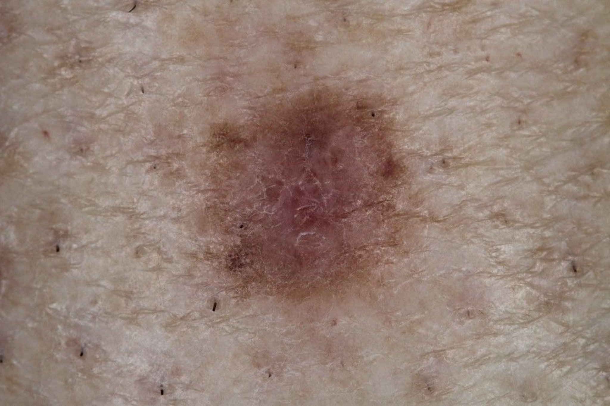
Dermatofibromas are firm, red-to-brown, small papules or nodules composed of fibroblastic tissue. They usually occur on the thighs or legs but can occur anywhere.
Topic Resources
Image courtesy of Marie Schreiner, PA-C.
Dermatofibromas are common among adults, more so in women. Their cause is probably genetic. Lesions are usually 0.5 to 1 cm in diameter, firm, and may dimple inward with gentle pinching. Most lesions are asymptomatic, but some itch or ulcerate after minor trauma.
Diagnosis of Dermatofibromas
Clinical evaluation
Diagnosis of dermatofibromas can often be made clinically. There are several described dermatoscopic patterns of dermatofibromas (1). Lesions are sometimes biopsied to exclude melanocytic proliferation (eg, nevus, solar lentigo, melanoma) or other tumors. Dermatofibromas may appear hyperpigmented in dark skin tones.
Diagnosis reference
1. Zaballos P, Puig S, Llambrich A, et al: Dermoscopy of dermatofibromas: A prospective morphological study of 412 cases. Arch Dermatol 144(1):75-83, 2008. doi: 10.1001/archdermatol.2007.8
Treatment of Dermatofibromas
Excision if troublesome
Dermatofibromas that cause troublesome symptoms can be excised. Treatment with cryosurgery may alleviate symptoms.

