


Topic Resources


Keloids are more frequent in patients with dark skin. Keloids are most likely to occur in people genetically prone to them, after skin injury or inflammation (including after surgery and burns), especially in areas susceptible to keloid formation (1). These areas include the earlobes, shoulders, upper arms, and anterior chest. Keloids are considered distinct from hypertrophic scars because keloidal scar tissue extends beyond the margins of the wound or injury and hypertrophic scar tissue does not. Keloids may also appear spontaneously. They may be raised as much as 0.5 centimeters or more above the epidermis.
Keloids are characterized histopathologically by excessive collagen and extracellular matrix deposition. Keloids are shiny, firm, smooth, usually ovoid but sometimes contracted or webbed, and slightly pink or hyperpigmented. They can be associated with pruritus and pain. Extensive keloids can cause significant cosmetic and functional impairment.

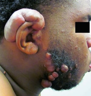
This photo shows keloids on the ear and beard area after dog-bite injuries.
© Springer Science+Business Media

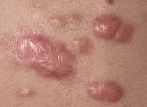
A keloid is hypertrophied tissue that develops in an area of injury or spontaneously; keloids are shiny, smooth, often dome-shaped, and slightly pink or hyperpigmented.
Image provided by Thomas Habif, MD.

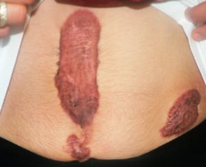
In distinction from hypertrophic scars, keloids extend beyond the borders of the original wound invading normal skin. The patient has multiple large keloid scars on the anterior abdominal wall, after intra-abdominal surgery.
© Springer Science+Business Media
This photo shows keloids on the ear and beard area after dog-bite injuries.
© Springer Science+Business Media
A keloid is hypertrophied tissue that develops in an area of injury or spontaneously; keloids are shiny, smooth, often dome-shaped, and slightly pink or hyperpigmented.
Image provided by Thomas Habif, MD.
In distinction from hypertrophic scars, keloids extend beyond the borders of the original wound invading normal skin. The patient has multiple large keloid scars on the anterior abdominal wall, after intra-abdominal surgery.
© Springer Science+Business Media
Reference
1. Limandjaja GC, Niessen FB, Scheper RJ, Gibbs S. The Keloid Disorder: Heterogeneity, Histopathology, Mechanisms and Models. Front Cell Dev Biol. 2020;8:360. Published 2020 May 26. doi:10.3389/fcell.2020.00360
Diagnosis of Keloids
History and physical examination alone
Diagnosis of keloids is clinical; a history of skin injury, along with the presence of characteristic hypertrophied and often hyperpigmented lesions in people with dark skin should raise suspicion for a keloid.
Treatment of Keloids
Possibly glucocorticoid injection, excision, gel sheeting, and/or immunomodulators
Treatment of keloids is often ineffective and recurrence is common.
Monthly glucocorticoid injections (eg, triamcinolone acetonide) into the lesion sometimes flatten the keloid. Monthly glucocorticoid injections (eg, triamcinolone acetonide) into the lesion sometimes flatten the keloid.
Surgical or laser excision may debulk lesions, but the risk is that they recur larger than before (1). Excision is more successful if preceded and followed by a series of intralesional glucocorticoid injections. Gel sheeting (applying a soft, semiocclusive dressing made of cross-linked polymethylsiloxane polymer, or silicone) or pressure garments are other adjunct therapies to prevent recurrence.
Immunomodulators (eg, topical imiquimod) have been used to prevent keloid development or recurrence. Immunomodulators (eg, topical imiquimod) have been used to prevent keloid development or recurrence.
Treatment references
1. Bailey J, Schwehr M, Beattie A. Management of Keloids and Hypertrophic Scars. Am Fam Physician. 2024;110(6):605-611.
Drugs Mentioned In This Article


