


- Angiostrongyliasis
- Anisakiasis
- Ascariasis
- Baylisascariasis
- Dracunculiasis
- Hookworm Infection
- Pinworm Infestation
- Strongyloidiasis
- Toxocariasis
- Trichinosis
- Trichuriasis
- Overview of Filarial Nematode Infections
- Bancroftian Lymphatic Filariasis and Brugian Lymphatic Filariasis
- Dirofilariasis
- Loiasis
- Mansonellosis
- Onchocerciasis (River Blindness)
Enterobiasis is an intestinal infestation by the pinworm (a type of nematode) Enterobius vermicularis. It usually occurs in school-aged children, but adult members of their household and caregivers, people who live in long-term care facilities, and those who have anal-oral contact with an infected partner during sex are also at risk. Its major symptom is perianal or vaginal itching. Diagnosis is by visual inspection for threadlike worms in the perianal area or the cellophane tape test for ova. Treatment is with mebendazole, pyrantel pamoate, or albendazole.. It usually occurs in school-aged children, but adult members of their household and caregivers, people who live in long-term care facilities, and those who have anal-oral contact with an infected partner during sex are also at risk. Its major symptom is perianal or vaginal itching. Diagnosis is by visual inspection for threadlike worms in the perianal area or the cellophane tape test for ova. Treatment is with mebendazole, pyrantel pamoate, or albendazole.

Pinworm infestation is estimated to be present in more than 1 billion people worldwide. Prevalence is highest among school-aged children (for example, one study reported a prevalence of approximately 20% of kindergarten and primary school children in Europe) (1).
Because most cases occur in school-aged and young children, their family members or caregivers are at increased risk of infection. Risk is also increased in people who live in long-term care facilities. Infection can be transmitted through anal-oral sexual contact with an infected partner.
(See also Approach to Parasitic Infections.)
General reference
1. Wendt S, Trawinski H, Schubert S, Rodloff AC, Mössner J, Lübbert C. The Diagnosis and Treatment of Pinworm Infection. Dtsch Arztebl Int. 2019;116(13):213-219. doi:10.3238/arztebl.2019.0213
Pathophysiology of Pinworm Infestation
Pinworm ova become infective within a few hours of reaching the perineum. Infestation usually results from transfer of ova from the perianal area to fomites (clothing, bedding, furniture, rugs, toys, toilet seats), from which the ova are picked up by the new host, transmitted to the mouth, and swallowed. Thumb sucking is a risk factor. Reinfestation (autoinfestation) easily occurs through finger transfer of ova from the perianal area to the mouth.
Pinworms reach maturity in the lower gastrointestinal tract within 2 to 6 weeks. The female worm migrates out of the anus to the perianal region (usually at night) to deposit ova. The sticky, gelatinous substance in which the ova are deposited and the movements of the female worm cause perianal pruritus. The ova can survive on fomites as long as 3 weeks at normal room temperature.

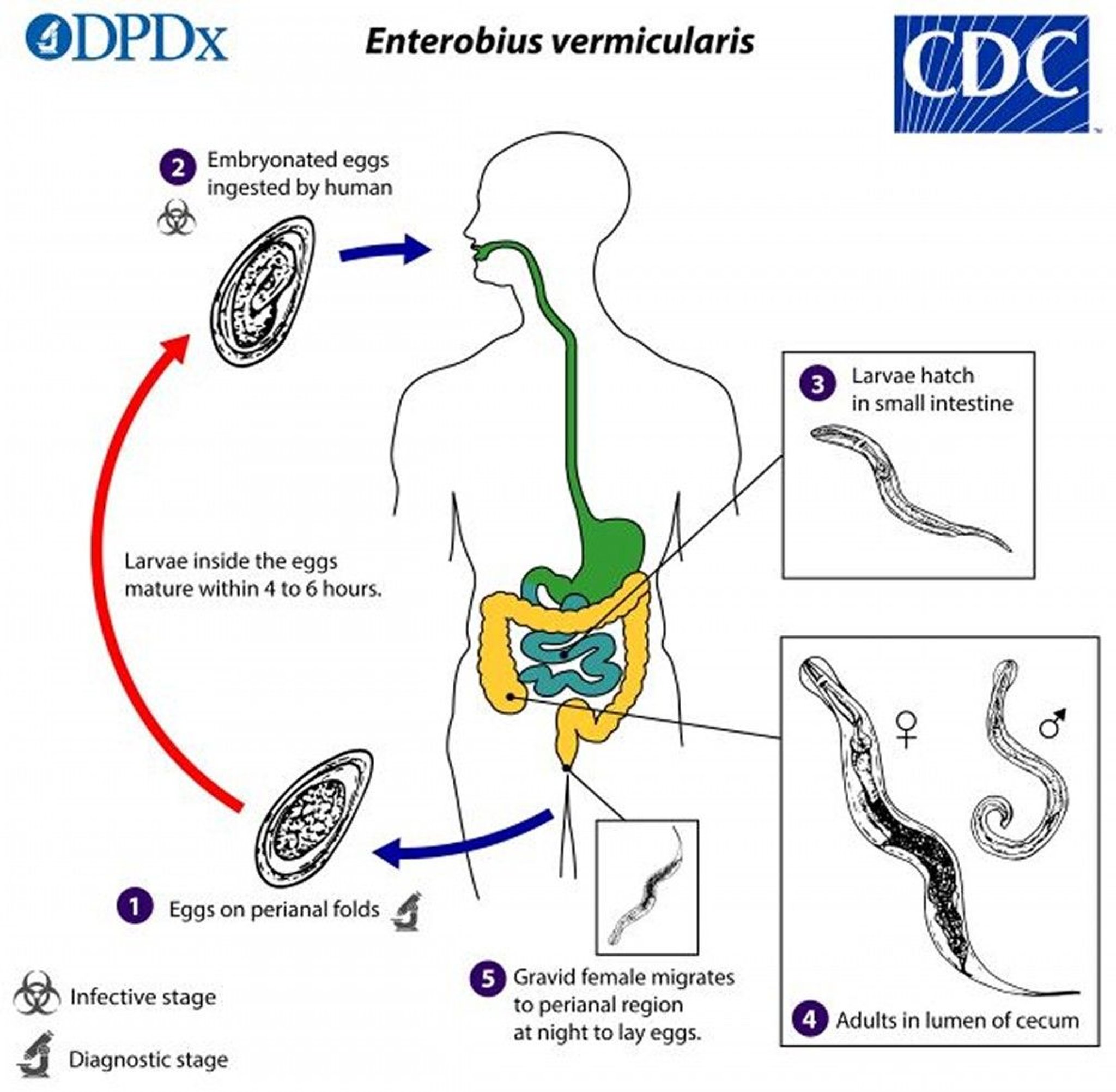
Image from the Centers for Disease Control and Prevention, Global Health, Division of Parasitic Diseases and Malaria.
Symptoms and Signs of Pinworm Infestation
Most infected people have no symptoms or signs, but some experience perianal pruritus and develop perianal excoriations from scratching. Secondary bacterial skin infection may occur. Pinworms sometimes migrate to the vagina, causing pruritus and vaginitis. Rarely, they ascend through the upper female genital tract, causing peritoneal lesions.
Many other conditions (eg, abdominal pain, insomnia, seizures) have been attributed to pinworm infestation, but a causal relationship is unlikely. Pinworms have been found obstructing the appendiceal lumen in cases of appendicitis, but the presence of the parasites may be coincidental.
Diagnosis of Pinworm Infestation
Examination of the perianal region for worms, ova, or both
Pinworm infestation can be diagnosed by finding the female worm, which is 8 to 13 mm long (males are 2 to 5 mm), in the perianal region 1 or 2 hours after going to bed at night or in the morning or by using a low-power microscope to identify ova on cellophane tape. Samples are obtained in the early morning before defecating or arising by patting the perianal skinfolds with a strip of cellophane tape, which is then placed sticky side down on a glass slide and viewed microscopically. The 50- by 30-micron ova are oval with a thin shell that contains a curled-up larva. A drop of toluene placed between tape and slide dissolves the adhesive and eliminates air bubbles under the tape, which can hamper identification of the ova. This procedure should be repeated on 3 successive mornings if necessary.
Similarly, if pinworm infestation is suspected in a child with vaginal pruritus or discharge (usually because there is a recent history of pinworm infection in the child or in a friend, classmate, or family member), a piece of cellophane tape can be applied to the vaginal introitus.
On occasion, the diagnosis can be made by examining samples taken from under the patient's fingernails.
Less frequently, eggs may also be detected in stool, urine, or vaginal specimens.
Treatment of Pinworm Infestation
Mebendazole, pyrantel pamoate, or albendazoleMebendazole, pyrantel pamoate, or albendazole
Because pinworm infestation is seldom harmful, prevalence is high, and reinfestation is common, treatment is indicated only for symptomatic infections. However, most parents actively seek treatment when their children have pinworms.
A single dose of mebendazole, pyrantel pamoate, or albendazole, repeated in 2 weeks, is effective in eradicating pinworms (but not ova) in A single dose of mebendazole, pyrantel pamoate, or albendazole, repeated in 2 weeks, is effective in eradicating pinworms (but not ova) in> 90% of cases.
Carbolated petrolatum (ie, containing carbolic acid) or other antipruritic creams or ointments applied to the perianal region may relieve itching.
Prevention of Pinworm Infestation
Pinworm reinfestation is common because viable ova may be excreted for 1 week after therapy, and ova deposited in the environment before therapy can survive 3 weeks. Multiple infestations within the household are common, and treatment of the entire family may be necessary.
The following can help prevent the spread of pinworms:
Washing the hands with soap and warm water after using the toilet, after changing diapers, and before handling food (the most successful way)
Frequently washing clothing, bedding, and toys
If people are infected, showering every morning to help remove eggs on the skin
Vacuuming the environment to try to eliminate eggs
Avoidance of oral-anal contact during sex
Key Points
Pinworm infestation is the most common helminthic infection in the United States; most cases occur in school-aged or younger children, but adults who care for children, family members of an infected child, people who live in long-term care facilities, and those who have oral-anal contact during sex are also at risk.
Pinworm infestation is seldom harmful, and reinfestation is common.
Ova deposited in the environment can survive 3 weeks.
Pinworm eggs may be ingested when people touch their mouth after they scratch their perianal area or after they handle contaminated clothes or other objects (eg, bed linens).
Most infected people have no symptoms or signs, but some have perianal pruritus.
Diagnose pinworm infestation by collecting ova in the morning on cellophane tape and using a low-power microscope to identify them; diagnosis can also be made by finding the female worm in the perianal region 1 or 2 hours after a person goes to bed at night.
If patients are symptomatic, treat with mebendazole, pyrantel pamoate, or albendazole.If patients are symptomatic, treat with mebendazole, pyrantel pamoate, or albendazole.
Drugs Mentioned In This Article


