


- Angiostrongyliasis
- Anisakiasis
- Ascariasis
- Baylisascariasis
- Dracunculiasis
- Hookworm Infection
- Pinworm Infestation
- Strongyloidiasis
- Toxocariasis
- Trichinosis
- Trichuriasis
- Overview of Filarial Nematode Infections
- Bancroftian Lymphatic Filariasis and Brugian Lymphatic Filariasis
- Dirofilariasis
- Loiasis
- Mansonellosis
- Onchocerciasis (River Blindness)
Angiostrongyliasis is infection with larvae of nematodes (worms) of the genus Angiostrongylus. Clinical presentation and diagnosis differ by species. A. cantonensis causes eosinophilic meningitis, with neurologic symptoms, neck stiffness, and sometimes fever or other systemic symptoms. Diagnosis is by lumbar puncture and PCR testing of cerebral spinal fluid and serology, and treatment includes corticosteroids and supportive care. A. costaricensis causes eosinophilic enteritis (abdominal angiostrongyliasis) with gastrointestinal symptoms and sometimes fever or other systemic symptoms. Diagnosis is by surgical biopsy of abdominal tissues, and treatment is supportive care. For both species, treatment with anthelmintics is either ineffective or deleterious.
Topic Resources

Angiostrongylus are parasites of rats (rat lungworms). Excreted larvae are taken up by intermediate hosts (land snails and slugs) and paratenic or transport hosts (hosts that are not required for the parasite's development but that can transmit infection to humans, such as certain land crabs, freshwater shrimp and prawns, frogs, and toads). Human infection is acquired by ingestion of raw or undercooked intermediate or transport hosts or of raw produce that contains a small snail or slug or part of one. Mucus trails from slugs and snails may contain small numbers of larvae but are not considered major sources of infection (1).

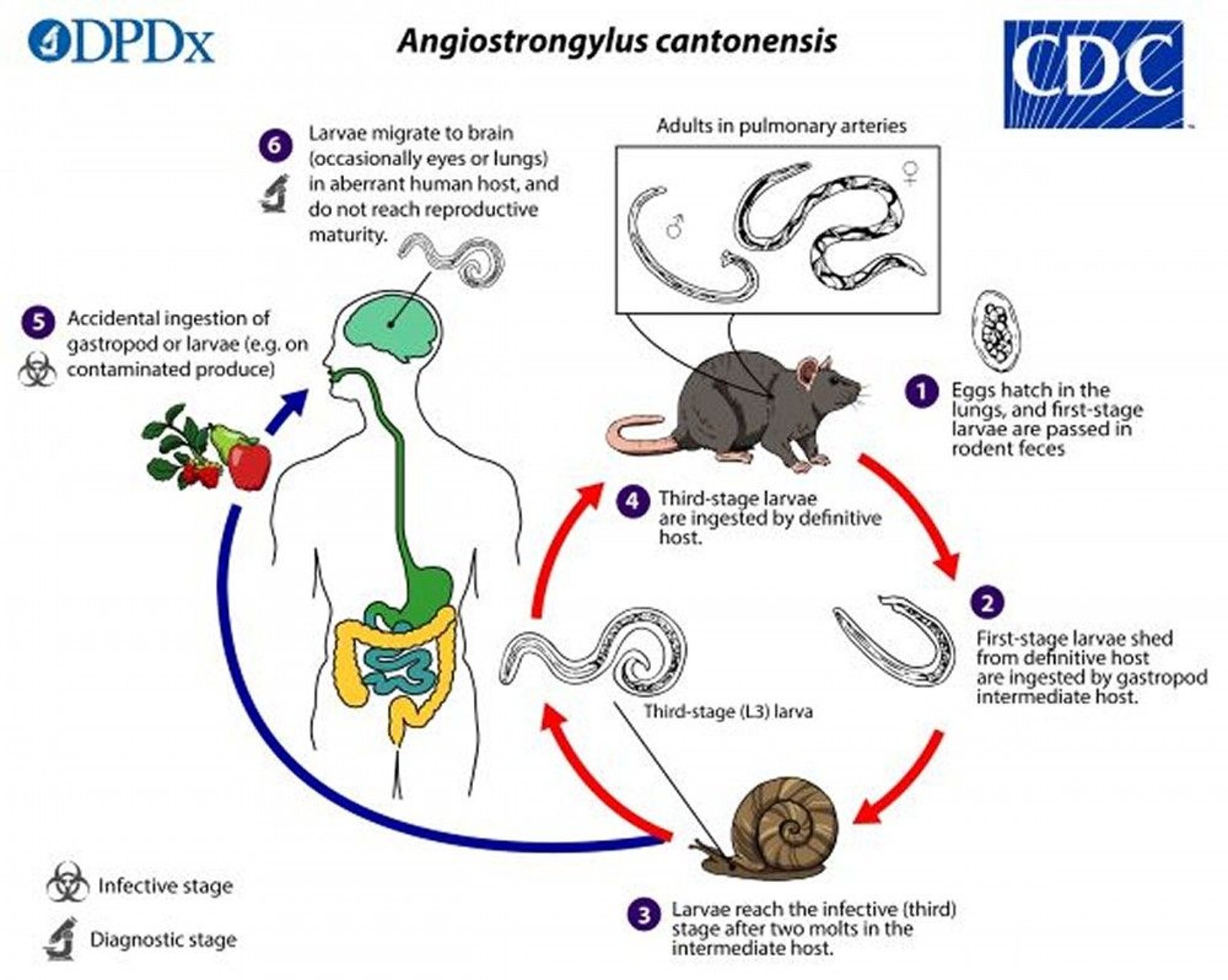
Image from the Centers for Disease Control and Prevention, Global Health, Division of Parasitic Diseases and Malaria.
A. cantonensis infection (neural angiostrongyliasis) occurs predominantly in Southeast Asia and the Pacific Basin, although infection has been reported elsewhere, including the Caribbean, Hawaii, and parts of the southern United States. It is one of the most common causes of eosinophilic meningitis worldwide (2). The larvae migrate from the gastrointestinal tract to the meninges, where they cause eosinophilic meningitis, with fever, headache, and meningismus, accompanied by eosinophilia. Occasionally, ocular invasion occurs.
A. costaricensis infection (abdominal angiostrongyliasis) occurs in the Americas, predominantly in Latin America and the Caribbean. Adult worms reside in arterioles of the ileocecal area, and eggs can be released into the intestinal tissues, resulting in local inflammation with abdominal pain, vomiting, and fever; this infection can mimic appendicitis. Abdominal angiostrongyliasis is often accompanied by eosinophilia, and a painful right lower quadrant mass may develop.
(See also Approach to Parasitic Infections.)
General references
1. Kramer KJ, Posner J, Gosnell WL. Role of Gastropod Mucus in the Transmission of Angiostrongylus cantonensis, a Potentially Serious Neurological Infection. ACS Chem Neurosci. 2018;9(4):629-632. doi:10.1021/acschemneuro.7b00491
2. CDC Yellow Book 2024: Angiostrongyliasis. Accessed November 27, 2024.
Symptoms and Signs of Angiostrongyliasis
Eosinophilic meningitis caused by A. cantonensis manifests with neurologic symptoms such as severe headache, diplopia, paresthesia, hyperesthesia, or seizures (1). Symptoms or signs of neck stiffness (nuchal rigidity) are typically present. Patients may develop a low-grade fever, body aches, fatigue, or gastrointestinal symptoms (eg, nausea, vomiting, abdominal pain). In severe cases, blindness, paralysis, or death can occur.
Eosinophilic enteritis caused by A. costaricensis manifests with abdominal pain, nausea, vomiting, and/or diarrhea and sometimes peritonitis or a palpable abdominal mass (2). Fever or other systemic symptoms (eg, fatigue) may be present. In severe cases, intestinal perforation may occur.
Symptoms and signs references
1. CDC Yellow Book 2024: Angiostrongyliasis. Accessed November 27, 2024.
2. Walger LK, Rodriguez R, Marcolongo-Pereira C, et al. Diagnostic criteria and case definitions for abdominal angiostrongyliasis: a systematic review from the Brazilian experience. Parasitol Res. 2024;123(3):155. Published 2024 Mar 6. doi:10.1007/s00436-024-08177-2
Diagnosis of Angiostrongyliasis
If meningitis is suspected (A. cantonensis), cerebrospinal fluid (CSF) analysis
If enteritis is suspected (A. costaricensis), surgical biopsy to identify eggs and larvae in various tissues or adult worms in the mesenteric arterial lumen or its branches
Complete blood count
Sometimes serology
Angiostrongyliasis is suspected based on a history of ingesting potentially contaminated material, including intermediate or transport hosts.
Patients with meningeal findings require lumbar puncture with PCR testing of CSF, which typically shows elevated CSF pressure, protein, and white blood cells with eosinophils > 10% (can be as high as 70%). Complete blood count shows eosinophilia > 5% in blood; A. cantonensis parasites are rarely visible. Peripheral eosinophilia may not always correlate well with CSF eosinophilia. Focal lesions are not usually seen on CT or MRI of the brain. A. cantonensis larvae and eggs are not present in stool.
Diagnosis of gastrointestinal infection due to A. costaricensis is difficult because larvae and eggs are not present in stool. Definitive diagnosis is made if abdominal surgery is performed (often for another suspected etiology of the symptoms, eg, appendicitis) and histologic evaluation identifies eggs or larvae in biopsies of various abdominal tissues or identifies adult worms in the mesenteric arterial lumen or its branches. A high percentage of eosinophils (>10%) may be present in blood and within infected tissue (1).
Immunoassays and molecular diagnostics are not widely available. The Centers for Disease Control and Prevention (CDC) offers a polymerase chain reaction (PCR) for A. cantonensis in CSF (2); molecular tests for A. costaricensis are available only in research laboratories.
Diagnosis references
1. Rojas A, Maldonado-Junior A, Mora J, et al. Abdominal angiostrongyliasis in the Americas: fifty years since the discovery of a new metastrongylid species, Angiostrongylus costaricensis. Parasit Vectors. 2021;14(1):374. Published 2021 Jul 22. doi:10.1186/s13071-021-04875-3
2. Qvarnstrom Y, da Silva AC, Teem JL, et al. Improved molecular detection of Angiostrongylus cantonensis in mollusks and other environmental samples with a species-specific internal transcribed spacer 1-based TaqMan assay. Appl Environ Microbiol. 2010;76(15):5287-5289. doi:10.1128/AEM.00546-10
Treatment of Angiostrongyliasis
For meningitis, corticosteroids and sometimes lumbar puncture to reduce intracranial pressure
A. cantonensis meningitis is treated with analgesics, removal of CSF at frequent intervals to reduce intracranial pressure; corticosteroids can decrease the frequency of therapeutic lumbar puncture (1). Most patients have a self-limited course and recover completely. Anthelmintic therapy may increase the inflammatory response because it results in the release of parasite antigens.
There is no specific treatment for A. costaricensis infection; most infections resolve spontaneously. Anthelmintics do not appear to be effective and may lead to additional migration of worms and worsening symptoms (2).
Treatment references
1. Ansdell V, Kramer KJ, McMillan JK, et al. Guidelines for the diagnosis and treatment of neuroangiostrongyliasis: updated recommendations. Parasitology. 2021;148(2):227-233. doi:10.1017/S0031182020001262
2. Loría-Cortés R, Lobo-Sanahuja JF. Clinical abdominal angiostrongylosis. A study of 116 children with intestinal eosinophilic granuloma caused by Angiostrongylus costaricensis. Am J Trop Med Hyg. 1980;29(4):538-544.
Prevention of Angiostrongyliasis
People who live in or travel to areas with A. cantonensis or A. costaricensis should avoid eating raw or undercooked snails, slugs, freshwater shrimp, land crabs, frogs, centipedes, and lizards as well as potentially contaminated vegetables and vegetable juices.
Key Points
Humans acquire Angiostrongylus when they consume raw or undercooked snails or slugs or transport hosts (certain land crabs, frogs, toads, or freshwater prawns or shrimp).
A. cantonensis larvae migrate from the gastrointestinal tract to the meninges, where they cause eosinophilic meningitis; A. costaricensis eggs can be released into the intestinal tissues, causing abdominal pain, vomiting, and fever.
Eggs are not present in the stool of patients with angiostrongyliasis.
Treat A. cantonensis neural infection with meningitis with analgesics, corticosteroids, and, if intracranial pressure is elevated, removal of cerebrospinal fluid at frequent intervals.
Treating A. costaricensis abdominal infection with anthelmintics does not appear to be effective and may lead to additional migration of worms and worsening symptoms.
Most Angiostrongylus infections resolve spontaneously.


